Medical expert of the article

New publications
Gonioscopy
Last reviewed: 07.07.2025

All iLive content is medically reviewed or fact checked to ensure as much factual accuracy as possible.
We have strict sourcing guidelines and only link to reputable media sites, academic research institutions and, whenever possible, medically peer reviewed studies. Note that the numbers in parentheses ([1], [2], etc.) are clickable links to these studies.
If you feel that any of our content is inaccurate, out-of-date, or otherwise questionable, please select it and press Ctrl + Enter.
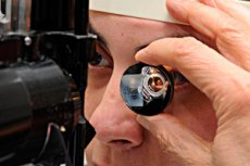
Gonioscopy is a method of examining the angle of the anterior chamber, hidden behind the translucent part of the cornea (limbus), which is performed using a gonioscope and a slit lamp.
During this examination, the patient's head is placed on the slit lamp stand, the chin and forehead are fixed, and the doctor first smears a special gel on the contact plane of the gonioscope and opens the eye slit of the patient's eye being examined with one hand, and with the free hand sets the contact plane of the gonioscope on the cornea of this eye. With one hand, the doctor holds the gonioscope, and with the other, using the handle of the slit lamp, moves the light slit along the edge of the gonioscope. The mirror plane of the gonioscope allows you to direct a beam of light into the corner of the anterior chamber of the eye and obtain a reflected image.
In medical practice, the most commonly used gonioscopes are Goldman (three-mirror cone-shaped), Van Beuningen (four-mirror pyramidal) and M. M. Krasnov (single-mirror). The gonioscope allows one to examine the distinctive features of the structure of the anterior chamber angle: the root of the iris, the anterior stripe of the ciliary body, the scleral spur to which the ciliary body is attached, the corneoscleral trabecula, the scleral venous sinus (Schlemm's canal), and the internal border ring of the cornea.
Determining the degree of openness of the anterior chamber angle is considered particularly relevant. According to the existing classification, the anterior chamber angle can be wide, medium-wide, narrow, and closed. With a wide angle, all its components are clearly visible, including the ciliary body strip and corneoscleral trabeculae. With an anterior chamber angle of medium width, the ciliary body is not visible or is defined as a narrow strip. If the anterior chamber angle is narrow, it is impossible to see either the ciliary body or the posterior part of the corneoscleral trabeculae. With a closed anterior chamber angle, the corneoscleral trabeculae are completely invisible, and the root of the iris is adjacent to the anterior border ring of Schwalbe.
Gonioscopy allows us to identify all sorts of pathological changes in the anterior chamber angle: goniosynechiae, newly formed vessels, tumors, foreign bodies.
 [
[