Medical expert of the article

New publications
Corneal biomicroscopy
Last reviewed: 07.07.2025

All iLive content is medically reviewed or fact checked to ensure as much factual accuracy as possible.
We have strict sourcing guidelines and only link to reputable media sites, academic research institutions and, whenever possible, medically peer reviewed studies. Note that the numbers in parentheses ([1], [2], etc.) are clickable links to these studies.
If you feel that any of our content is inaccurate, out-of-date, or otherwise questionable, please select it and press Ctrl + Enter.
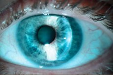
Corneal biomicroscopy is performed to systematically identify clinical signs, determine the location, depth and size of corneal damage.
Direct illumination method with diffuse light
Used to detect gross changes.
- A narrow oblique light slit allows examination of each quadrant of the cornea.
- Additional narrowing of the light beam allows visualization of very fine optical details.
- The change in the height of the coaxial beam is used to measure the extent of damage.
- The direction of the light slit can be changed by rotating the lamp housing.
- When the beam passes through all layers of the cornea, the thickness and depth of its damage are determined.
- The character of the light can be changed by using filters. With a red-free filter, red objects appear black, which increases the contrast of the image when examining vascular structures and when staining with rose bengal. A cobalt blue filter is used when staining with fluorescein.
Scleral scattering method
The light slit is decentered so that the light falls on the limbus, with the microscope focused in the center. The light is distributed inside the cornea due to total internal reflection and reaches the opposite limbus. The damaged area of the cornea is illuminated by the scattering of the light beam reflected in the thickness of the cornea. This method is important in determining subtle changes in the cornea.
Reflected light examination method
Using light reflected from the iris or fundus, it can detect subtle changes in the endothelium and epithelium, corneal precipitates, and small blood vessels.
What do need to examine?