Medical expert of the article

New publications
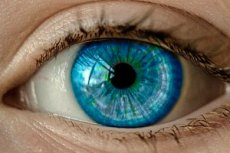
Corneal scraping and biopsy
Last reviewed: 07.07.2025

All iLive content is medically reviewed or fact checked to ensure as much factual accuracy as possible.
We have strict sourcing guidelines and only link to reputable media sites, academic research institutions and, whenever possible, medically peer reviewed studies. Note that the numbers in parentheses ([1], [2], etc.) are clickable links to these studies.
If you feel that any of our content is inaccurate, out-of-date, or otherwise questionable, please select it and press Ctrl + Enter.
The cornea is scraped with a Kimura spatula, a curved needle tip (for the hypodermis) or a blade. After instillation of a local anesthetic without preservatives, the edges and bottom of the lesion (usually an ulcer) are carefully and thoroughly scraped under slit lamp control. Contact lenses should also be examined.
The corneal material is placed on a glass slide for Gram staining and on appropriate media:
- blood agar (for most bacteria and fungi);
- thioglycollate broth (for most bacteria);
- chocolate agar (for Neisseria and Haemophilus);
- Sabouraud agar (for fungi); incubated at about 37 C;
- meat-peptone concentrated broth (for fungi that do not grow on Sabouraud agar);
- non-nutrient agar on plates inoculated with E. coli culture (for Acanthamoeba);
- yeast extract buffer agar (for Acanthamoeba).
NB: Media should be kept at room temperature before sowing.
Corneal biopsy is performed using a trephine or by open layer-by-layer dissection with a sharp blade.
Indications for corneal biopsy
- Keratitis with negative or inconclusive results of scraping and culture on media.
- Deep corneal infiltrate, the nature of which cannot be determined by simple scraping.
- Difficulties in diagnosis in corneal dystrophies or rare genetically determined storage diseases with corneal pathology.
Who to contact?