Medical expert of the article

New publications
Crystalline
Last reviewed: 04.07.2025

All iLive content is medically reviewed or fact checked to ensure as much factual accuracy as possible.
We have strict sourcing guidelines and only link to reputable media sites, academic research institutions and, whenever possible, medically peer reviewed studies. Note that the numbers in parentheses ([1], [2], etc.) are clickable links to these studies.
If you feel that any of our content is inaccurate, out-of-date, or otherwise questionable, please select it and press Ctrl + Enter.
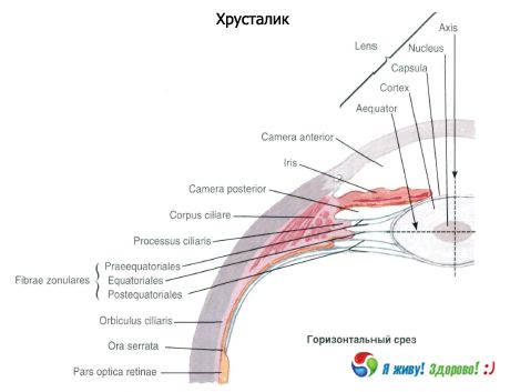
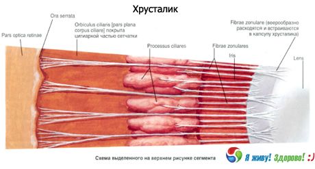
The lens is a transparent, light-refracting body that has the shape of a biconvex lens and is located in the eye between the iris and the vitreous body. After the cornea, the lens is the second refractive medium of the eye's optical system.
The anterior surface of the lens (facies anterior lentis) and its most prominent point, the anterior pole (polus anterior), face the posterior chamber of the eyeball. The more convex posterior surface (facies posterior) and the posterior pole of the lens (polus posterior lentis) are adjacent to the anterior surface of the vitreous body. The imaginary line connecting the anterior and posterior poles of the lens, which is on average 4 mm long, is called the axis of the lens (axis lentis). This axis coincides with the optical axis of the eyeball. The rounded peripheral edge of the lens, where its anterior and posterior surfaces converge, is called the equator. The substance of the lens (substantia lentis) is colorless, transparent, dense, and does not contain vessels or nerves. The inner part - the nucleus of the lens (nucleus lentis) is significantly denser than the peripheral part - the cortex of the lens (cortex lentis).
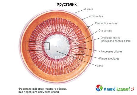
The lens is covered on the outside by a thin transparent elastic capsule (capsula lentis), which is attached to the ciliary body by means of the ciliary belt (Zinn's ligament), which extends from the lens capsule. The lens capsule is a structureless, glassy, elastic shell. The lens capsule has selective permeability, as a result of which the chemical composition of the transparent lens is stable.
When the ciliary muscle contracts, the choroid itself shifts forward, the ciliary body approaches the equator of the lens, the ciliary belt weakens and the lens seems to straighten. In this case, the anteroposterior size of the lens increases, it becomes more convex, its refractive power increases - the lens is set for close vision. In the case of relaxation of the ciliary muscle, the ciliary body moves away from the equator of the lens, the ciliary belt stretches, the lens flattens, its refractive power decreases and the lens is set for distant vision. The ability of the lens to see at different distances is called accommodation. Therefore, the lens together with the ciliary muscle (ciliary body) and the fibers connecting them are called the accommodative apparatus of the eye.
In young people, the lens fibers are soft and elastic. When the ciliary muscle contracts and the Zinn ligament relaxes, the lens takes on a more spherical shape, thereby increasing its refractive power. As the lens grows, the centrally located older lens fibers lose water, become denser, and become thinner, forming a dense lens core. This process, which prevents excessive enlargement of the lens (due to which the lens grows throughout life without increasing in size), begins very early, and by the age of 40-45, a well-formed dense core is already present. The lens fibers surrounding the core form the cortical layer of the lens. With age, due to the enlargement of the core and the reduction of the cortical layer, the lens becomes less elastic, and its accommodative capacity decreases. Metabolic processes in the lens occur extremely slowly. The exchange is carried out with the participation of the epithelial cells of the anterior capsule of the lens. They receive all the necessary substances from the intraocular fluid, which surrounds the lens on all sides.
The lens resembles a lentil in appearance. The curvature of the anterior surface is 10 mm, the posterior surface is 6 mm, i.e. the posterior surface is more convex, the thickness of the lens (diameter) is 9-10 mm. The lens weighs 0.2 g. In a child, the lens has a spherical shape. Identification zones:
- anterior and posterior pole - the centers of the anterior and posterior surfaces;
- axis - a line connecting the poles;
- equator - the line where the front surface transitions to the back.
Histological structure of the lens (capsule, epithelium, fibers, nucleus):
- capsule - a collagen-like membrane, part of which (zocular plate) can be separated from the anterior surface. The capsule is thicker in front;
- epithelium - these are hexagonal cells under the anterior capsule, which are retracted in the equatorial region;
- The fibers of the lens are hexagonal prisms. There are about 2.5 thousand fibers in total. Shifting toward the center, they grow toward the poles, but they do not reach the poles. Sutures are formed at the junctions of the anterior and posterior fibers with the capsule;
- nucleus - embryonic and adult. There are sutures in the embryonic nucleus. The adult nucleus, which is formed by the compaction of the lens fibers, is formed by the age of 25. The lens contains the following substances: water, proteins, mineral salts, lipids, ascorbic acid. The lens contains 60% water, 18% soluble proteins (alpha, beta and gamma proteins). The main protein - cysteine - ensures the transparency of the lens. 17% are insoluble proteins (albuminoids), which are contained in the membranes of the fibers; 2% - mineral salts, a small amount of fats.
 [
[ What's bothering you?
What do need to examine?
How to examine?