Medical expert of the article

New publications
Extra mammary glands under the arm
Last reviewed: 04.07.2025

All iLive content is medically reviewed or fact checked to ensure as much factual accuracy as possible.
We have strict sourcing guidelines and only link to reputable media sites, academic research institutions and, whenever possible, medically peer reviewed studies. Note that the numbers in parentheses ([1], [2], etc.) are clickable links to these studies.
If you feel that any of our content is inaccurate, out-of-date, or otherwise questionable, please select it and press Ctrl + Enter.
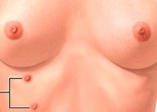
Additional mammary glands under the armpit are an anomaly. The fact is that the extra lobules are located directly in the armpit area. Not all women know about their little peculiarity. This is usually discovered at a doctor's appointment. Sometimes, the lobes are noticeable even with the naked eye. In some cases, the milky stream opens directly into the armpit. Visually, this resembles a common pimple. During pregnancy and childbirth, this anomaly becomes obvious.
 [ 1 ], [ 2 ], [ 3 ], [ 4 ], [ 5 ], [ 6 ], [ 7 ], [ 8 ], [ 9 ], [ 10 ]
Causes additional mammary glands under the arm
The reasons for the appearance of this anomaly have not yet been fully determined. More precisely, there is still no consensus regarding the factors that contribute to their development.
Usually, additional lobes of the mammary glands appear due to genetic disorders. This happens against the background of a hormonal imbalance or surge. For example, this could be the same menopause, pregnancy or puberty.
Accessory glands have always been considered pathologies. In fact, this is an anomaly. A woman's breasts are symmetrically located. Naturally, they consist of only 2 glands. Accessory
"growth" can be located under normal glands. But, there are more "interesting" places of their presence. Thus, most often the anomaly develops on the neck, armpits and back. Very rarely, an additional organ can appear on the genitals.
Often the reason lies in the incorrect development of the mammary gland rudiments. This happens due to problems with development. This process begins as early as the 6th week of development. Usually by the 10th week all the extra "growths" are eliminated on their own, as a result there are only two mammary glands left in the place where they should be. But, in some cases, elimination never occurs.
Symptoms additional mammary glands under the arm
The symptoms are purely visual. But it is worth noting that this pathology can also be painful. In some cases, a woman develops a complex. After all, this affects her psychological state, and brings a lot of inconvenience. This leads to the emergence of fears and dislike for her own body.
Additional organs are characterized by a voluminous form. In some cases, they have a nipple. Sometimes the neoplasm has a special form or resembles a normal mammary gland. Usually, its location is in the armpit area.
A few days before the onset of menstruation, the anomaly can change significantly in volume. Before menstruation, the breast often increases in size. As for the additional organ, it follows its example. During pregnancy and after childbirth, it can even secrete breast milk.
This anomaly is not oncology. But, it is not worth excluding the possibility of its "transformation" into malignancy. Such cases have actually occurred. The danger of developing oncology remains in those people whose additional organ is constantly injured, for example, by clothing.
Additional breast lobe under the arm
A quite usual habitat for this anomaly is an additional lobe. Sometimes, the location is in other areas, but this is the most pronounced. It is worth noting that not in all cases this mammary gland has anything in common with the main one.
Under the arm, a neoplasm occurs in almost 6% of all cases. The growth takes its development from the embryonic rudiments. This process lasts throughout the entire milk line. In total, there are eight types of anomalies. Half of them do not contain glandular tissue. But despite this, they have a fully functional nipple. Experts do not consider these anomalies to be life-threatening. Yes, this is a neoplasm, but not malignant and it cannot develop into this form. However, this issue has not been thoroughly studied.
Women with an additional gland agree to surgery. They make this decision because of constant discomfort. The presence of this organ weighs them down and causes a number of complexes. After all, in many cases, the additional gland interferes with normal existence and creates a number of inconveniences.
When examining an anomaly on an X-ray image, a low-intensity zone can be seen, usually it is darkened. It is mainly surrounded by special fibers.
Diagnostics additional mammary glands under the arm
Diagnostics includes several basic methods. The visual method allows you to examine the breast for anomalies. It is easy to do. Sometimes, the nipple is poorly developed, and it can easily be confused with a mole. If the patient is plump, then the additional lobe is differentiated from a lipoma or cyst.
Additional laboratory tests, as well as instrumental methods, are prescribed if there is a suspicion of the development of a pathological process. This examination is also carried out before prescribing high-quality treatment for an anomaly. You can start with a consultation with a mammologist. If there are gynecological disorders, you will also have to visit a gynecologist, you can go to an appointment with a surgeon - gynecologist.
Some studies help to assess the main features of the glands, what functions they perform, whether there are any inflammatory processes in them. Such methods include ultrasound, computed tomography and MRI.
Ultrasound is the highest quality and most effective diagnostic method. This method allows you to detect the development of neoplasms that are both on the surface and deep under the skin. In addition, it is ultrasound that allows you to find significant differences between different formations in organs. This could be a cyst or mastitis. The study should be carried out exclusively in the first phase of the cycle.
Computer tomography is a method of obtaining an X-ray image. Thanks to it, the specialist not only receives a high-quality image, but also sees the state of the organ layer by layer. This allows you to identify all the small details and see possible pathologies.
MRI of the mammary glands is a method somewhat similar to computed tomography. However, in this case, X-ray radiation is not used. Evaluation of this study is necessary for making a decision regarding further treatment, including surgical intervention.
What do need to examine?
Who to contact?
Treatment additional mammary glands under the arm
Treatment can only be surgical. It is not necessary to remove the neoplasm surgically. It is not dangerous. But women decide to have surgery because the neoplasm prevents them from living a full life. Basically, psychological problems arise. There is a desire to remove the uncomfortable neoplasm.
Plastic surgery with complete removal of the "growth" is recommended only if there is a strong cosmetic defect. The reason for removal may be constant pain, as well as impaired functionality. Heredity may be an indication for the use of a surgical method. In this case, we mean the presence of malignant breast tumors in one of the relatives.
Surgical intervention is a correction by liposuction. That is, the neoplasm is simply "sucked out". If this method is not possible, the growth is removed and the skin tissue is sutured. The method completely depends on the size and structure of the accessory gland. If the formation is large and consists partly of fatty tissue, then an incision of no more than 5 mm is made and a layer of fat is pumped out. Sometimes the incision is made much larger. In some cases, it is necessary to remove the skin over the abnormal gland.
The surgery lasts about an hour. The patient can be sent home the same day, since local anesthesia is used. The stitches are removed a week after the removal. There are no special recommendations regarding behavior after the surgery.
After the surgery, a scar remains, but since it is in the armpit area, it is not visible. Therefore, it will not create any inconvenience. After the operation, the woman can live a full life.
Prevention
Prevention of additional mammary glands under the armpit is impossible in principle. It is not possible to control this process or do anything to prevent it. After all, as mentioned above, this is a common anomaly. Nothing influences its development. The only factor may be hormonal imbalance or heredity. It is impossible to do anything about it.
Adolescence, the beginning of the menstrual cycle, pregnancy - all this occurs with the participation of a large number of hormones. It is impossible to prevent these processes, unless the last one. Therefore, the only way to get rid of the problem is to simply remove the neoplasm.
There is nothing scary or dangerous in this procedure. The operation is simply performed and that's it. Then the woman will be able to live without much discomfort. If there is no desire, you can leave everything as is. But, it is worth repeating once again, it is impossible to prevent the appearance of this neoplasm.
Forecast
The prognosis of additional mammary glands under the arm depends entirely on whether the woman is bothered by it or not. In any case, it is not worth leaving the anomaly unattended. Any defect can lead to negative consequences.
If the patient does not want to resort to removal, she should be regularly examined by a doctor. For this, a visual examination is carried out and an ultrasound is performed. This will allow monitoring the growth and structure of the abnormal formation.
It is necessary to remove additional organs if they are subject to constant trauma. This occurs due to clothing, pressing with limbs, etc. Constant trauma can lead to a malignant neoplasm, which will entail a number of complications.
Once everything is removed, a favorable course can be predicted. The occurrence of this anomaly cannot be called rare. It is important that the problem is correctly diagnosed, and the degree of danger is adequately assessed. After which the question of further treatment with surgical removal is decided.