
Osteophyte of the heel bone
Last reviewed: 07.06.2024

All iLive content is medically reviewed or fact checked to ensure as much factual accuracy as possible.
We have strict sourcing guidelines and only link to reputable media sites, academic research institutions and, whenever possible, medically peer reviewed studies. Note that the numbers in parentheses ([1], [2], etc.) are clickable links to these studies.
If you feel that any of our content is inaccurate, out-of-date, or otherwise questionable, please select it and press Ctrl + Enter.
Known to many heel spur is nothing but an osteophyte of the heel bone. It is a spiky or awl-shaped growth of bone tissue, which is formed as a result of inflammatory diseases, moderate and severe traumatic injuries, degenerative-dystrophic processes that contribute to changes in the structure of the bone.
Epidemiology
Osteophyte of the heel bone is an important orthopedic problem, which is associated with severe pain syndrome, limitation of motor functions. Often, patients with calcaneal osteophytes temporarily lose the ability to work, lose the ability to lead an active lifestyle, engage in sports.
Pathologic growths can be found in people of different age groups, but they are most often found in people over 45 years of age, and especially in patients with overweight, rheumatoid arthritis, and diabetes mellitus. In the young and middle-aged population, the prevalence of PCS is 11-21%. This rate is consistent across nationalities: 11% in India, 13% in Ireland, 15% in Zimbabwe, 16% in Thailand, 17% in Europe, and 21% in the Americas. [1], [2] This rate increases with age to 55% in those over 62 years of age, to 59-78% in those with current or previous heel pain, and to 81% in those with osteoarthritis. [3], [4] This problem often accompanies other pathologies or curvatures of the foot that may require surgical treatment. [5]
Osteophyte of the medial tuberosity of the heel bone was first identified and described by the German Dr. Plettner back in 1900. At that time, he coined the term "heel spur".
Osteophytes are diagnosed and treated by orthopedic trauma doctors.
Causes of the osteophyte of the heel bone
Osteophytes of the heel bone appear as a result of metabolic disorders, trauma to the heel, excessive loads on the bone.
The most common causative factors are considered to be:
- Inflammatory reactions; [6]
- Degenerative processes (heel spurs commonly occur in all arthritis, with estimates of up to 80% in osteoarthritis and 72% in rheumatologic patients over 61 years of age); [7], [8]
- Fractures;
- Prolonged forced leg positions; [9]
- Bone neoplasms;
- Endocrine pathologies (obesity);
- Flat feet, other foot deformities.
Depending on the cause of occurrence, osteophytes of the heel bone are:
- Degenerative-dystrophic (associated with impaired blood circulation and trophism in the area of the heel bone);
- Post-traumatic (as a consequence of a fracture, contusion);
- Tumorigenic (caused by malignant neoplasms);
- Endocrine (related to hormonal disorders);
- Neurogenic (as a result of damage to the peripheral or central nervous system).
In many cases, the appearance of osteophytes of the heel bone is associated with pathologies such as arthrosis and arthritis.
The majority of patients with osteophytes are elderly and old people. In them, the appearance of the problem is most often associated with degenerative changes. As for children and young people, the situation is different: osteophytes appear mainly due to infectious or autoimmune processes.
Risk factors
The factors that could contribute to heel bone osteophytes are not fully understood. Among the most likely:
- Frequent mechanical damage to bones and ligaments (excessive body weight [10] and overloading, improperly fitted shoes, etc.);
- Metabolic disorders causing degenerative changes in the fascia;
Rubin & Witten (1963 ) found that 46% of patients with calcaneal osteophytes were overweight compared to 27% of controls, and Moroney et al (2014 ) found that 82% of people with calcaneal osteophytes were overweight or obese. Moreover, after adjusting for age and gender, people with calcaneal osteophytes were 6.9 times more likely to be obese compared to those without calcaneal steophytes (Menz et al. 2008title="Plantar calcaneal spurs in older people: longitudinal traction or vertical compression? - PMC">).
Due to the constant inflammatory process in the plantar fascia, the flexible ligamentous tissue is replaced by bone tissue - that is, tissue ossification occurs. The formed bony overgrowth leads to permanent damage to the soft tissue structures of the sole, developing plantar fasciitis. Osteophytes of the heel bone are present in 45-85% of patients with plantar fasciitis; they also have a number of common risk factors such as obesity and advanced age, suggesting that these two factors may be related etiologically. [11], [12]
Among the possible triggering factors:
- Longitudinal type of flatfoot;
- Hypodynamia, overloading the foot;
- Obesity;
- Prolonged static overload, prolonged standing, wearing unsuitable and/or uncomfortable shoes;
- Frequent mechanical injuries to the feet (in particular, during active sports).
Pathogenesis
Osteophyte of the heel bone is a pathological outgrowth, often single, sometimes multiple. The shape can vary from serrated or spiky to massive and bumpy. The structure of the osteophyte does not differ from normal bone tissue.
Osteophytes happen:
- Bone-compact;
- Bone-spongy;
- Bone and cartilage;
- Metaplastic.
Bone-compact osteophytes are formed from the compact substance of bone tissue, one of the types of tissue that make up bone. This substance performs many functions, it is very strong and mechanically resistant, and it "stores" the main necessary chemical elements - in particular, phosphorus and calcium.
Bone spongy osteophytes are formed from spongy tissue, which has a cellular structure and is formed from bone membranes and plates. This substance is light and not particularly strong.
Bone and cartilage osteophytes appear as a result of deformation of cartilage in the area of articular surfaces, which can be associated with overloading of the joint, inflammatory and degenerative pathologies.
The appearance of metaplastic osteophytes is due to the replacement of one type of cell in the bone tissue by another - for example, due to inflammatory or infectious processes, as well as impaired bone regeneration.
Symptoms of the osteophyte of the heel bone
The most obvious sign of a calcaneal osteophyte is considered to be severe pain during walking - and especially when taking the first steps ("starting pain") after a long break or rest. As the calcaneal osteophyte develops and enlarges, the pain becomes more intense. [13]
The immediate onset of pain syndrome does not always indicate that the abscess is already present. In many patients, pain appears long before the formation of osteophyte, and from the moment of development of the inflammatory process in the soft tissues of the heel and destruction of the plantar fascia.
Osteophyte of the plantar surface of the heel bone can give rise to pain of varying intensity, which depends on the stage of the inflammatory reaction and the degree of damage to the fascia. Often the pain is acute: it feels as if a sharp spike has been thrust into the heel. [14], [15]
Massive osteophytes of the heel bone can lead to shortening of the plantar fascia. At the same time, it is weakened and the foot is curved. The gait changes, which is caused by severe pain and the inability to fully support the heel (patients try to step on the toe or the outside of the foot).
Osteophyte of the calcaneal tuberosity is accompanied by pain syndrome in the posterior part of the ankle joint, with irradiation to the fingers of the affected limb, the muscles of the lower leg. Pain tends to intensify in the afternoon or after prolonged stay "on the feet".
Beak osteophyte of the heel bone can be accompanied by edema, which is due to inflammatory reaction, microcirculatory disorders, direct destruction of tissues.
Among the main symptoms are:
- Redness, lividity of the skin in the area of the heel;
- The appearance of calluses, corns;
- Pressure and burning sensation, increased sensitivity and tingling in the heel area;
- Limp.
As the pathological formation grows, the symptoms worsen after prolonged loading of the lower limbs. Osteophyte of the right heel bone often makes itself known with a sharp support on the heel (for example, at the time of a sharp rise from a chair or sofa), as well as when climbing stairs. Less often, the pathology proceeds with only a slight discomfort, but this happens only in isolated cases.
Osteophyte of the left heel bone is accompanied by an obvious gait disturbance. The patient tries to put the affected foot in such a way as not to touch the sore spot, relying mainly on the toes and the back of the foot. In many patients, such manipulations lead to the development of left-sided transverse flat feet.
With intensive growth of the bone neoplasm, especially in its awl-shaped form, a fracture of the osteophyte of the heel bone is not excluded. In this case, the patient's ability to move independently is almost completely lost, which is associated with the appearance of unbearable pain when loading the foot. [16]
Where does it hurt?
Complications and consequences
Patients suffering from osteophytes of the heel bone are forced to limp, change the position of the foot, stepping on the toes with a transition to the lateral part of the foot. This can lead to the following complications:
- Curvature of the foot and ankle;
- Swelling and pain in the lower leg;
- Arthritis and arthrosis affecting the ankle joint and the joint of the big toe;
- Flat feet (development of a deformity or aggravation of a pre-existing problem);
- Spinal curvature.
If the osteophyte grows to a significant size, a fracture may occur (complete or partial, in the form of a bone fracture). In such a situation, the patient completely loses the ability to step on the affected limb, which negatively affects the quality of life.
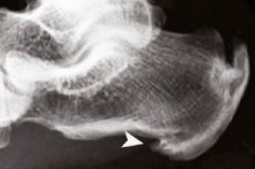
Diagnostics of the osteophyte of the heel bone
Diagnostic appointments are made by an orthopedic doctor. First of all, it is necessary to perform radiography to visualize the state of the bone apparatus, configuration of the bones, their location and size.
Among the ancillary diagnostic procedures:
- General and blood biochemical study, assessment of the probability of inflammatory reaction development, determination of uric acid indices in the blood;
- General urinalysis;
- Ultrasound examination of the affected area to assess the condition of soft tissue structures, detection of possible purulent foci;
- Examination of the vascular apparatus of the lower extremities to detect a possible circulatory disorder;
- Magnetic resonance examination of the foot to assess structural condition.
On individual indications may require consultations with specialists of narrow profile: endocrinologists, traumatologists, vascular surgeons, oncologists and others.
What do need to examine?
Differential diagnosis
Pain in the area of the heel bone is not always due to the formation of an osteophyte. A similar picture may accompany:
- Gout;
- Osteomyelitis;
- Rheumatoid arthritis;
- Bone tuberculosis;
- Bechterew's disease;
- Partial and complete fractures of the heel bone, soft tissue injuries;
- Foot deformities.
You should not practice self-medication and take analgesics and anti-inflammatory drugs on your own. Treatment is prescribed by a doctor based on the results of diagnosis and final diagnosis.
Who to contact?
Treatment of the osteophyte of the heel bone
To get rid of osteophyte of the heel bone, a comprehensive approach is used. Treatment is supervised by an orthopedic surgeon, traumatologist or surgeon.
It is important to minimize the physical load from the affected foot. For this purpose, the patient is selected orthopedic shoes, insoles, special wrist inserts.
Drug treatment is aimed at eliminating the inflammatory response. Nonsteroidal anti-inflammatory drugs (oral preparations, as well as ointments, gels, creams) are indicated.
Additionally prescribe massage, physiotherapy (electrophoresis, hydrotherapy) to optimize metabolic processes and eliminate inflammation.
If the usual conservative methods do not bring relief, medication blockade is performed by injecting the affected heel with injectable solutions of analgesics - in particular, Diprospan. This method is effective, but it is not recommended to use it often, due to the increased risk of destruction of ligaments and fascia.
Particularly effective is considered shockwave treatment - a special physiotherapeutic technique, which consists in the application of low-frequency acoustic-impact oscillations. Thanks to this treatment:
- Optimizes blood and lymph circulation;
- Metabolic processes at the local level are improved;
- Relaxes the spasmed muscles;
- Stops the development of the inflammatory process;
- Relieve pain, repair damaged tissue.
A course of shockwave treatment usually consists of 6-8 sessions. Its effectiveness is estimated at about 97%. However, this procedure has its own contraindications:
- During pregnancy;
- Presence of oncologic diseases, acute infectious processes;
- The presence of a pacemaker;
- High blood pressure;
- Impaired blood clotting;
- Vascular inflammation, venous thrombosis;
- Childhood (including adolescents).
Rarely, in particularly severe cases, surgical treatment is prescribed, which consists of removing the bone growth. The affected limb is fixed with a plaster cast, which is removed approximately four weeks after the completion of rehabilitation measures.
More information of the treatment
Prevention
The appearance of osteophytes can be prevented, as well as slow down the development of existing small growths, if you competently adjust lifestyle and follow these recommendations of experts:
- Choose only high-quality and comfortable shoes with a small comfortable heel height of no more than 3-4 cm;
- If possible, use special unloading orthopedic insoles with supinator;
- Control your own weight, prevent the development of obesity;
- Eat a well-balanced diet and drink sufficient fluids throughout the day;
- Maintain adequate physical activity, take frequent walks, and avoid overloading the feet with prolonged standing or heavy ("impact") loads;
- Massage the feet regularly;
- Watch your posture, do exercises to prevent deformities of the spine and feet.
If the first signs of discomfort in the heel area are detected, it is necessary to visit an orthopedist. Most conservative therapies are most effective just at the early stages of osteophyte development and allow you to stop further progression of pathological growths.
Forecast
The prognosis of the disease depends on the intensity of growth of osteophytes, as well as on the timeliness and competence of treatment. If pain or discomfort appears in the heel area, it is important not to delay to visit a doctor, a qualified orthopedist, who will prescribe diagnostic and appropriate therapeutic measures. The following therapeutic manipulations may be required:
- Pain blockades;
- Physiotherapy;
- Therapeutic massage, physical therapy.
In addition, the doctor prescribes drug therapy in accordance with modern approaches, with mandatory monitoring of effectiveness.
Some patients prefer to self-treat, use various folk methods. However, it is important to realize that you can not completely get rid of the problem, so it is better to consult a specialist beforehand. Osteophyte of the heel bone is a disease with a combined etiology, so it is necessary to affect it in a variety of ways, using both medicines for ingestion and external, including physiotherapeutic, effects.