
Left ventricular aneurysm
Last reviewed: 07.06.2024

All iLive content is medically reviewed or fact checked to ensure as much factual accuracy as possible.
We have strict sourcing guidelines and only link to reputable media sites, academic research institutions and, whenever possible, medically peer reviewed studies. Note that the numbers in parentheses ([1], [2], etc.) are clickable links to these studies.
If you feel that any of our content is inaccurate, out-of-date, or otherwise questionable, please select it and press Ctrl + Enter.
Aneurysm of the left ventricle of the heart (ventriculus sinister cordis), from which the great circle of blood circulation begins, is a blood-filled localized fibrous bulge arising in the area of the weakened wall of this heart structure.
Epidemiology
More than 95% of left ventricular aneurysms are caused by myocardial infarction and coronary heart disease; left ventricular aneurysm after infarction is statistically reported in 30-35% of cases.
At least one third of cases are associated with congenital anomalies of the heart and coronary vessels. Congenital left ventricular aneurysms (most often asymptomatic) diagnosed for the first time in adults are rare. They are diagnosed in adults after 40 years of age with a prevalence of 0.3-04% of cases.
Ventricular heart aneurysms in children are very rare. [1]
Causes of the left ventricular aneurysms
As a rule, damage to the heart wall with the formation of its bulging zone, which changes the shape of the ventricle and negatively affects its function, is caused by transmural, i.e. Full-layer myocardial infarction - involving all layers (epicardium, myocardium and endocardium). In such cases, a postinfarction left ventricular aneurysm is defined. [2]
In addition, the causes of this cardiovascular pathology may be related to:
- Coronary heart disease (CHD);
- Isolated systolic arterial hypertension;
- Inflammation of the heart muscle - myocarditis;
- Trauma or heart surgery;
- Degeneration or myocardial degeneration dystrophy of various etiologies.
Left ventricular aneurysms can also result from congenital/genetic defects including:
- Left ventricular hypertrophy;
- Aortic valve dysfunction (between the left ventricle and the aorta) leading to chronic aortic insufficiency;
- Mitral valve prolapse and tricuspid (tricuspid) valve dysplasia;
- Open artioventricular canal;
- Coronary anomalies in the form of left coronary artery branching from the pulmonary artery with intracardiac shunting between the circulatory circles.
Also read - acute and chronic cardiac aneurysms: ventricular, septal, postinfarction, congenital
Risk factors
In addition to acute myocardial ischemia, heart failure and the previously named congenital defects, experts consider risk factors for the formation of left-sided ventricular aneurysm:
- Coronary circulation problems due to atherosclerosis and occlusion of arterial vessels of the heart;
- Elevated BP - arterial hypertension;
- Dilated cardiomyopathy, in which the inner part of the left ventricular myocardium has a spongy structure (so-called noncompact myocardium);
- A history of tuberculosis or rheumatism (rheumatic fever);
- Sarcoidosis, often resulting in left ventricular wall thinning and cavity dilatation, as well as cardiac amyloidosis and vasculitis;
- Increased production of thyroid hormones (hyperthyroidism), which affect the overall hemodynamics and can cause thyrotoxic cardiomyopathy with myocardial damage, dilatation of the heart chambers and left ventricular hypertrophy.
And athletes should be aware that long-term use of anabolic steroids increases the development of coronary atherosclerosis and damage to the ventricular myocardium. [3]
Pathogenesis
The mechanism of congenital ventricular aneurysm formation is presumably related to abnormalities during ontogenesis (embryonic formation) of the heart, which subsequently lead to an increase in ventricular volume. Intrauterine ischemic myocardial injury and endocardial fibroelastosis - with fibrous tissue overgrowth causing abnormal heart enlargement and ventriculus sinister cordis hypertrophy - are also not excluded.
As for the acquired aneurysm of this localization, its pathogenesis as a complication of myocardial infarction is the most studied.
After infarction, part of the myocardium of the ventricular wall as a result of acute ischemia is damaged or undergoes necrosis with death of cardiomyocytes (because in adults, cardiac muscle cells have left the active phase of the cell cycle and practical lost the ability to reproductive mitosis and regeneration).
In this case, the damaged myocardium is replaced by fibrous tissue, and the area formed in the ventricular wall becomes not only thinner - with reduced strength, but also inert. That is, this area does not participate in the contraction of the heart muscle even during systole (ventricular contraction to push blood out of the heart into the systemic bloodstream) and gradually expands, bulging outside the ventricular wall. [4]
Symptoms of the left ventricular aneurysms
Most left ventricular aneurysms are asymptomatic and are detected incidentally on echocardiographic examination. [5]
The general clinical picture is determined not only by the size of the aneurysm and its shape, but also by the volume of intact (functioning) wall tissue, and consists of left ventricular insufficiency of varying degrees, the symptoms of which are manifested:
- Shortness of breath (on exertion and at rest);
- Rapid fatigue, dizziness and fainting;
- A feeling of heaviness behind the sternum and pain radiating to the left shoulder and shoulder blade - angina pectoris;
- Sustained ventricular (ventricular) tachyarrhythmia - a disturbance in the rhythm of systolic ventricular contractions with their increase in frequency;
- Wheezing on inhalation, noisy breathing;
- Swelling of the feet.
Forms
There is no single unified classification of left ventricular aneurysms, but aneurysms are divided into congenital and acquired aneurysms according to their origin.
Some specialists among the acquired pathologies distinguish ischemic or postinfarction - left ventricular aneurysms after infarction; traumatic (after cardiac surgery); infectious (formed in patients with infective endocarditis, rheumocarditis, polyarteritis nodosa, tuberculosis, etc.), as well as idiopathic (of unknown etiology).
Postinfarction ventricular aneurysms are divided into acute and chronic aneurysms. An acute left ventricular aneurysm forms within two days (maximum two weeks) after myocardial infarction, while a chronic left ventricular aneurysm forms within six to eight weeks.
The localization of the pathological bulge is also taken into account. Apical left ventricular aneurysm - left ventricular apex aneurysm - is a bulge in the anterior part of the upper segment of the left ventricular wall. It accounts for one-third to one-half of all cases, and the first signs are manifested by ventricular tachyarrhythmias.
Left ventricular anterior wall aneurysms form in approximately 10% of cases; left ventricular posterior wall aneurysms are diagnosed in 23% of patients; inferior posterior wall aneurysms account for no more than 5% and lateral wall aneurysms for 1% of cases.
Submitral (subvalvular) annular left ventricular aneurysm is a rare cardiac pathology and can occur after infarction, in congenital posterior mitral valve defect, endocarditis, or rheumocarditis.
Aneurysms are also classified according to their shape. While a sac-shaped aneurysm is characterized by a rounded thin-walled bulge of the ventricular wall (consisting of myocardium with varying degrees of fibrous replacement) and the presence of a narrowed "entrance" part (neck), a diffuse aneurysm of the left ventricle has a wider communication with the ventricular cavity and therefore looks flatter when visualized. [6]
Complications and consequences
Accompanied by significant symptoms, left ventricular aneurysms can produce complications and cause sequelae, including:
- General decrease in systolic and diastolic heart function and development of secondary congestive heart failure;
- Blood stasis-related thrombosis - a wall thrombus in a left ventricular aneurysm that can dislodge and threaten to embolize, for example, the brain with the risk of subsequent stroke;
- Aneurysm rupture with cardiac tamponade.
Diagnostics of the left ventricular aneurysms
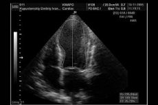
The diagnosis of left ventricular cardiac aneurysm is established by imaging studies, and clinical instrumental diagnosis uses ECG, Echocardiography (two or three-dimensional transthoracic echocardiography), chest radiography, MRI, computed tomographic coronary angiography, and several other instrumental methods of cardiac investigation.
Basic blood tests include: general, biochemical, for C-reactive protein, for troponin, alkaline phosphatase and creatine kinase levels.
Differential diagnosis
Differential diagnosis is very important because such aneurysms can mimic angina pectoris, Takotsubo cardiomyopathy, pericarditis/myocarditis, etc.
A true aneurysm must be differentiated from a pseudoaneurysm. While a true aneurysm is formed by a full thickness bulge of the ventricular wall, a false left ventricular aneurysm is formed by a rupture of the ventricular wall enclosed in the surrounding pericardium. Pseudoaneurysms are most often localized in the posterior and inferior walls of the left ventricle. [7]
Who to contact?
Treatment of the left ventricular aneurysms
Treatment methods for left ventricular aneurysms are determined based on clinical presentation and patient-specific data. Small to medium-sized aneurysms without symptoms can be safely managed with an expected five-year survival rate of up to 90%.
Drug treatment is aimed at reducing the intensity of symptoms and preventing complications. Medications of such pharmacological groups as:
- Cardiotonic cardiac glycosides - celanide (Lanatoside C) and others;
- Diuretics (diuretics), and aldosterone receptor antagonists - verospiron (Spironolactone) or inspra (Eplerenone);
- Beta-adrenoblockers - vasocardin (Corvitol), carvedilol, Propranolol, alotendine and other antiarrhythmic drugs;
- Anticoagulants (Warfarin ) - to prevent thromboembolism (during the first three months after a heart attack) and thrombolytics - Aspirin, Clopidogrel (Plavix or diloxol ), etc.;
- ACE (angiotensin-converting enzyme) inhibitors - Lisinopril, captopril, Perindopril, etc.
Surgical treatment should be performed in patients with left ventricular aneurysms with large bulge size; worsening cardiac function (chronic heart failure), significant ventricular arrhythmias, lateral thrombus formation with risk of embolism, and associated complications with risk of rupture.
The surgery that involves excising the aneurysm and placing a dacron patch on the ventricular wall is called Dore plasty or endoventricular circular plasty (EVCPP). [8]
Prevention
Experts believe that the incidence of aneurysm development, formed as a complication of myocardial infarction, can be reduced by early - in the acute phase of the disease - the resumption of blood supply (revascularization) damaged ischemic heart muscle tissue and, possibly, the use of ACE inhibitors.
Forecast
Large symptomatic left ventricular aneurysms can cause sudden cardiac death: within three months after infarction, the mortality rate is 67%, and after one year it reaches 80%. And compared to a heart attack without an aneurysm, mortality within a year is more than six times higher in patients with postinfarction aneurysms.
Long-term prognosis in symptomatic postinfarction aneurysms is largely determined by the level of left ventricular function before surgical intervention and the success of surgical treatment.
Some reports have shown that patients whose primary disability was related to angina pectoris and heart/ventricular failure have a five-year postoperative survival rate of 75-86%.