
Epiphyseolysis of the tibia
Last reviewed: 07.06.2024

All iLive content is medically reviewed or fact checked to ensure as much factual accuracy as possible.
We have strict sourcing guidelines and only link to reputable media sites, academic research institutions and, whenever possible, medically peer reviewed studies. Note that the numbers in parentheses ([1], [2], etc.) are clickable links to these studies.
If you feel that any of our content is inaccurate, out-of-date, or otherwise questionable, please select it and press Ctrl + Enter.
Damage to the epiphyseal cartilage or epiphyseal plate at the junction of the metaphysis and epiphysis of the tibia - with separation (detachment) of cartilage tissue - is defined as epiphyseolysis of the tibia. [1]
Epidemiology
It is known that growth plate fractures and epiphyseolysis are twice as common in boys as in girls because girls stop growing earlier and most of them have their growth plates transformed into mineralized bone tissue by 13-15 years of age (and boys by 15-18).
According to clinical statistics, after the distal radius of the forearm, the distal tibia is the second most common site of growth plate fracture. Almost half of the cases are associated with a Salter-Harris type II tibial fracture, where the fracture line passes through the bone body and exits through the metaphysis.
Injuries to the proximal tibial epiphysis are rare (0.5-3% of all cases), and this is because this epiphysis is protected by the ligaments of the knee.
Causes of the epiphyseolysis of the tibia.
The epiphysis is the thickened end of tubular bones, and the metaphysis adjacent to the epiphyseal plate (lamina epiphysialis) is the part of the bone where longitudinal growth occurs due to epiphyseal hyaline cartilage. Epiphyseolysis of the tibia is a pathology of the immature skeleton, because by the age of 14-17 years, epiphyseal closure occurs, that is, ossification of the growth plate. In adults, only a rudimentary epiphyseal line remains in its place.
Orthopedists attribute the causes of epiphyseolysis of the tibia to epiphyseal fractures of its proximal (upper) or distal (lower) portion.
Because of the increased shear and bending stresses in young adults, there are special forms of bone fracture, Salter-Harris fractures of several types, which involve the growth plates and damage them by forming a gap that disrupts the structure and function of the epiphyseal cartilage in the process of endochondral ossification.
Thus, distal tibial epiphyseolysis in most cases is the result of type IV fractures that cross the bone body almost vertically, extending from the metaphysis to the epiphysis. In such cases, the medial (inner) ankle is involved, with the fracture extending to the lower metaphysis of the tibia.
And epiphyseolysis of the tibial tuberosity (tuberositas tibiae) can result from a fracture of the upper tibia - in the proximal region of the tibia.
Detachment of the cartilage plate is also accompanied by the so-called Tiyo fracture, a fracture of the anterolateral epiphysis of the tibia, which is usually observed in adolescents with external trauma to the foot with rotation relative to the tibia.
In addition, epiphyseolysis of this bone may be seen in inversion and crush injuries of the upper and lower tibia.
Read also - bone and joint injuries in children
Risk factors
In addition to childhood and adolescence, fractures and obesity, experts note risk factors somehow associated with damage and possible detachment of epiphyseal cartilage such as:
- Fibrotic ostitis of post-traumatic or infectious origin;
- Lesions of bone tissue and periosteum of infectious-inflammatory nature - osteomyelitis;
- The destruction of the tibial tuberosity and the diaphyseal nucleus of its ossification caused by overloading (repeated stress injuries) of the lower limbs - in the form of schlatter's osteochondropathy;
- Metaphyseal dysostosis (dysplasia) in the form of the rare genetic Pyle's disease - with thickening of the ends of long bones and narrowing of their diaphysis, which increases the likelihood of fractures.
In addition, there is an increased risk of fractures, including shin bones at:
- Degenerative and dystrophic changes in bone tissue;
- Secondary hyperparathyroidism, because excessive production of PTH (paratgormone) not only reduces bone mineral density, but also activates osteoclasts, causing bone resorption and erosive tissue lesions of the epiphyses of tubular bones;
- Hypocalcemia, associated with vitamin D deficiency in the body or renal insufficiency and hyperphosphatemia.
Children with various neuromuscular disorders and myopathic syndrome. Are at risk for bone fractures and epiphyseal dislocation.
Pathogenesis
In explaining the pathogenesis of this acute osteochondral injury in children and adolescents, experts point out that the growth plates are the softest and weakest parts of the immature skeleton and have a very specific structure.
In fracture, fibrotic changes occur in the area connecting the epiphysis and metaphysis of the bone: the chondrocytes of the growth cartilage columns lose their intercellular connections and are partially replaced by connective tissue, which shifts under shear stress.
In fractures of types I-II - with horizontal and oblique splitting of the epiphyseal zone - there may be microscopic cracking of the epiphyseal plate, which separates the cell tables in the longitudinal direction. As a result of type III fractures (with splitting of the cancellous bone tissue of the epiphysis with deviation towards the epiphyseal plate), a part of the growth cartilage may completely move away from its place.
Also read - bone Development and Growth
Symptoms of the epiphyseolysis of the tibia.
The stages of growth plate displacement are defined as mild (displacement angle ˂ 30°), moderate (30-50°), and severe (at ˃ 50° displacement).
The first signs are manifested by localized fever, the appearance of swelling and hematoma at the end of the bone - near the knee joint or ankle (depending on the location of the tibial injury).
Clinical symptoms of a growth plate fracture may include pain and soreness, especially in response to pressure on the growth area; inability to move the affected limb and/or transfer body weight to it, i.e., exert downward pressure. To varying degrees, range of motion is limited and difficulty walking is experienced.
Complications and consequences
The main complications and consequences of this injury of the distal epiphysis are associated with premature partial closure of the bone growth zones and cessation of endochondral ossification, i.e. Longitudinal growth of the tibia, leading to limb asymmetry - their different lengths, which is accompanied by lameness.
These complications also occur in proximal tibial epiphysiolysis, but they are less common. And the younger the child is at the time of injury, the more likely it is to develop shortening and angular deformity, as the proximal tibial epiphysis grows about 6 mm per year until maturity.
In cases of epiphysiolysis due to vertical fracture of the epiphysis and metaphysis, there is often frontal or sagittal displacement of the injured limb with the development of arthritis.
Blount's disease, a disease of the upper (proximal) metaphysis of the tibia, which is a progressively increasing deformity of the tibia with outward curvature, internal tibial torsion, and pathologic changes in the knee joint, may also develop.
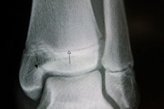
Diagnostics of the epiphyseolysis of the tibia.
This osteochondral lesion can be detected by instrumental diagnostics, including X-ray of the lower leg bones (both limbs), arthrography (X-ray of the intercostal, knee and ankle joints in two projections), and osteoscintigraphy. CT and MRI are also used for diagnosis, allowing visualization of soft tissues.
Differential diagnosis
A differential diagnosis with aseptic necrosis of bone and periosteum, joint tuberculosis, osteogenic sarcoma, dissecting osteochondritis, etc. Is performed.
Who to contact?
Treatment of the epiphyseolysis of the tibia.
For growth plate fractures, treatment depends on its severity. Less severe fractures usually only require a plaster cast or splinting.
But when the epiphyseal fracture crosses the growth plate or enters the joint and is poorly aligned, surgical treatment with percutaneous epiphyseodesis/osteosynthesis with transphyseal screws or tibial osteotomy and rigid fixation with an internal plate may be required.
After this intervention, x-rays should be taken periodically (for several years while the patient grows) to monitor the condition of the epiphyseal cartilage.
With proper treatment, most growth plate fractures heal without complications.
More details in the publication - fractures
Prevention
Only prevention of fractures and treatment of diseases that increase their risk can prevent tibial epiphysiolysis.
Forecast
If left untreated, the child or teen may become disabled.