
Right ventricular aneurysm
Last reviewed: 07.06.2024

All iLive content is medically reviewed or fact checked to ensure as much factual accuracy as possible.
We have strict sourcing guidelines and only link to reputable media sites, academic research institutions and, whenever possible, medically peer reviewed studies. Note that the numbers in parentheses ([1], [2], etc.) are clickable links to these studies.
If you feel that any of our content is inaccurate, out-of-date, or otherwise questionable, please select it and press Ctrl + Enter.
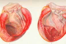
An extremely rare pathology, a right ventricular aneurysm, is a limited protrusion of the thinning and non-contractile right ventricular wall consisting of dead or scar tissue. It is more often a complication of a massive infarction or a consequence of congenital heart disease. [1]
Epidemiology
One of the primary causes of right ventricular aneurysm formation experts call ischemic heart disease, ischemia and right ventricular myocardial infarction. Limited right ventricular myocardial infarction is rare, as it is diagnosed in only 4% of patients who die from a heart attack. Much more common is right ventricular infarction arising on the background of transmural infarction of the left ventricle with inferior localization and posterior part of the interventricular septum. Approximately every third patient with inferior infarction has right ventricular damage.
Often, and extensive infarction entails the development of a right ventricular aneurysm. The problem can be detected in patients with chronic heart failure, as well as in postinfarction survivors (about a year after the attack).
Right ventricular aneurysm develops more often in men than in women (approximately 6 times). The pathology is diagnosed both in middle-aged and elderly patients. Congenital right ventricular aneurysms occur in only a few cases. [2]
Causes of the right ventricular aneurysms
Causes of possible right ventricular aneurysm development include volume overload, exposure to excessive resistance, tricuspid and pulmonary regurgitation, cardiomyopathy, etc.
In some congenital heart defects, there is a reduction of the right ventricular cavity, which is due to underdevelopment of the inflow or trabecular compartments, or hypertrophic processes in the heart muscle in the trabecular ventricular compartment.
Right ventricular aneurysm as a separate pathology is extremely rare. The problem is more often combined with other disorders, such as severe stenosis or atresia of the pulmonary artery, Fallot's tetrad, atresia of the tricuspid valve, and so on.
Among the possible causes of a right ventricular aneurysm are:
- Increased blood pressure;
- Hypertrophic form of cardiomyopathy;
- Amyloidosis;
- The final stage of aortic stenosis;
- Mitral stenosis;
- Pulmonary atresia or stenosis;
- "blue" heart defect (Fallot's tetrad ).
All these pathologic conditions can occur with right ventricular activity or with hypokinetic reduction of its functional capabilities and with the development of heart failure. [3]
Risk factors
Normal contractile function of the heart is associated with maximally efficient ejection against the background of minimal energy expenditure. This mechanism of cardiac muscle contractility is called synergy. Aneurysm of the right ventricle due to replacement of muscle tissue with scar tissue or due to impaired blood supply leads to a violation of this synergy: akinesia (non-participation in contractions of part of the ventricular wall) and dyskinesia (paradoxical pulsation) occurs. The quality of contractile function directly depends on the volume and location of the pathologic bulge, as well as on the preserved functionality of the healthy portion of the heart muscle.
The main factor in the development of right ventricular aneurysm is an extensive infarction accompanied by the formation of a thin scar. The larger the area of the lesion, the larger the area with scar tissue becomes. Under the influence of pressure inside the ventricle, the scar begins to bulge, and an aneurysm is formed. Additional factors can become:
- Physical activity in the acute phase of myocardial infarction, failure to comply with strict bed rest in the postinfarction period;
- High blood pressure;
- Congenital heart defects.
Aneurysm of the right ventricle can be formed both in the acute phase of infarction and at a remote stage, several months, a year after the attack. Occurrence of the bulge in the acute period occurs at the stage of myomalacia, and in the remote period is associated with weakness of the fibrous scar. [4]
It is possible to form a right ventricular aneurysm after cardiac surgery. Possible causes include:
- Pericardectomy;
- Focal myocardial shock;
- Damage with subsequent ischemia associated with inadequate myocardial protection of the right ventricle during surgery;
- Mechanical trauma to the heart tissue.
Pathogenesis
Chronic right ventricular aneurysm develops more often than the acute form of pathology. Usually its development is caused by chronic pulmonary hypertension.
Aneurysm is accompanied by a deterioration of contractility, which is characteristic of heart muscle damage: such is possible in right ventricular myocardial infarction, cardiomyopathy, as well as after cardiac surgery.
Left ventricular aneurysms are much more common, with increased pressure in the small circle of circulation and common interventricular mechanisms able to cause a problem on the right ventricular side.
Aneurysm of the right ventricle may develop against the background of volume loading, tricuspidal or pulmonary regurgitation, congenital heart disease. The sources of tricuspid regurgitation are:
- Tricuspid valve pathologies (rheumatism, congenital developmental defects, carcinoids, myxomatous degenerations, connective tissue dysplasia, etc.);
- Pathologic conditions of the right ventricle and small circle of circulation.
The causes of pulmonary regurgitation are predominantly:
- Increased pulmonary artery pressure;
- Surgical interventions for congenital heart disease (in the long term).
Macroscopy distinguishes these varieties of right ventricular aneurysm:
- Diffuse - represented by bulging of a limited area of scar tissue, with gradual flow to an area of normal muscle tissue.
- Mesenteric - characterized by the presence of a "neck" with its gradual expansion and the formation of a significant sized mesenteric cavity.
- A dissecting aneurysm is caused by endocardial rupture and is characterized by the appearance of a cavity in the muscle under the epicardium. When such an aneurysm ruptures, blood escapes into the pericardial cavity limited by adhesions, resulting in the formation of a false aneurysm.
In some patients, multiple cardiac aneurysms are diagnosed, as well as additional ("daughter") bulges from the aneurysmal wall.
About one in two patients has aneurysmal cavity thrombosis, but most cases are small wall thrombi. [5]
Symptoms of the right ventricular aneurysms
Symptomatology in right ventricular aneurysm is nonspecific and manifested by a general disturbance of cardiac activity. In particular, the patient may pay attention to the following symptoms:
- Cardiac, chest pains;
- Discomfort in the heart area after stressful or physical activity;
- Difficulty breathing, tachycardia;
- Dizziness, intermittent blackouts;
- Swelling of the extremities;
- Sensation of shortness of breath (especially often in the form of nocturnal attacks).
The first signs of right ventricular damage can be directly related to the disorder of the function of the great circle of blood circulation. Initially, the disease is asymptomatic, but many patients develop it:
- Ascites;
- Liver enlargement;
- Cyanosis of the nasolabial triangle area;
- Skin pallor;
- Unsubstantiated dyspepsia in the form of loose stools, nausea, bloating, etc;
- Instability in blood pressure readings.
Often, right ventricular aneurysm manifests itself against the background of the underlying disease, which entails an intertwining of symptoms. In this regard, it is possible to make a correct diagnosis only after a thorough and comprehensive diagnosis, with the involvement of medical specialists from different areas.
Aneurysm of the right ventricular pulmonary arterial trunk
A pulmonary artery aneurysm is said when the patient has a painful-limited expansion of the vascular trunk, which comes out of the right ventricle. In most cases, pathology is not accompanied by pronounced symptomatology, only some patients have intrathoracic pain, hoarseness, nocturnal and exertional dyspnea, hemoptysis. The disease is diagnosed based on the indicators of functional and radiological studies (chest X-ray, angiopulmonography), computer and magnetic resonance imaging of pulmonary vessels.
This aneurysm is quite rare: it is found mainly in patients over 50 years old, and most often accidentally, as in about 80% of cases the disease is not accompanied by any distinct and specific symptomatology.
The presumed cause of the development of the pathologic process is a congenital defect of a certain zone in the wall of the pulmonary arterial trunk. With increasing pressure in the small circle of circulation, this defect worsens, and the vascular wall stretches and thins. In the aneurysm cavity there is turbulence of blood flow, hemodynamic processes in the distal network of vessels are disturbed. Subsequently, the pressure on the stretched tissues increases, degenerative-dystrophic processes increase, there is a risk of rupture of the aneurysm wall. In many cases, the deposition and calcification of thrombi inside the cavity is noted.
Treatment of the pathology is surgical: the dilation is excised, or the vessel segment is resected with further prosthesis, or the aneurysm wall is reinforced with a lavsan prosthesis. Wait-and-see (observation) tactics are appropriate only in relation to small asymptomatic aneurysms.
Complications and consequences
Over time, pathologic dilation with bulging of the right ventricular wall may progress. The damaged tissue thins, loses elasticity and density. The aneurysm ruptures or stratifies, massive bleeding or parenchymatous hemorrhage develops. Infarct pneumonia may develop.
If the membranes of the aneurysm rupture, which communicates with the bronchial lumen, there is intrapulmonary bleeding. If the rupture occurs in the pericardial cavity, cardiac tamponade develops.
When thrombotic elements are detached and transported with blood from the aneurysm cavity, the risk of vascular thrombosis is significantly increased.
Specialists point out the following variants of adverse effects of right ventricular aneurysm:
- Enlargement and change in the shape of the right ventricle, with increased intramuscular tension of intact myocardium, increased oxygen demand of the heart muscle, and increasing picture of heart failure;
- Blood stasis in the small circulation;
- Clot formation, thromboembolic complications;
- Severe arrhythmias;
- Myocardial infarction (including recurrent), fatal.
Diagnostics of the right ventricular aneurysms
In most cases, if a right ventricular aneurysm is suspected, diagnostic measures involve not only a cardiologist, but also a vascular surgeon and a pulmonologist. During the initial examination, if possible, the primary and background pathology is detected, auscultation and percussion are performed. To make a final diagnosis, instrumental diagnostics is appointed:
- Cardiodiagnostic measures: electrocardiography allows to reveal the picture of overloaded right heart, the presence of bulging right ventricle. When echocardiography (cardiac ultrasound) is performed, hemodynamic disorders, valve insufficiency, arterial and wall dilatations are noted.
- Radiography: demonstrates the presence of a rounded mass in the right ventricle. Peripheral vascular abnormalities are represented by multiple or single compacted rounded shadows. To clarify the individual moments of pathology, angiopulmonography is performed.
- Computed tomography, magnetic resonance imaging is used to clarify the localization of the right ventricular aneurysm, its size and wall thickness. Tomographic methods are considered more accurate than similar radiologic procedures.
Tests can help to diagnose heart failure:
- A general blood test (iron deficiency anemia can be detected);
- General urinalysis (possible detection of cylindruria, proteinuria, indicating impaired renal function against the background of chronic heart failure);
- Blood biochemical study: AST, ALT, bilirubin and total protein, lactate dehydrogenase with creatine phosphokinase and MB fractions, myoglobin and electrolytes, cholesterol and C-reactive protein, coagulogram and BNP - level of brain natriuretic peptide.
Differential diagnosis
Right ventricular aneurysm should be differentiated with these pathologies:
- Disorders of the valve system of the heart;
- Myocarditis, cardiomyopathies;
- Right ventricular hypertrophy;
- Arterial stenosis with right ventricular hypertrophy;
- Hypertrophy due to amyloidosis;
- Ischemic heart disease with compensatory septal hypertrophy;
- Heart and lung tumors;
- Diaphragmatic hernia;
- Echinococcal cyst, coelomic pericardial cyst;
- Abdominomediastinal lipoma.
Who to contact?
Treatment of the right ventricular aneurysms
Conservative methods can not get rid of the right ventricular aneurysm, so when the first symptoms of heart failure appear, the doctor raises the question of surgical intervention. So, the main method of treatment of pathology is surgical excision of the problem area with subsequent suturing of the wall defect. Some patients additionally strengthen the aneurysmal wall with the help of polymer inserts.
Preoperative period includes medical preparation: if indicated, anticoagulants, cardiac glycosides, hypotensive drugs, oxygen therapy, oxygenobarotherapy. Strictly limited motor activity, exclude the influence of stress.
A cardiologist may prescribe the following medications as part of a recommended treatment regimen:
- Magnicor - antithrombotic drug - is taken in the amount of 75-150 mg per day, long-term. In some cases, digestive disorders, abdominal pain, nasal and gingival bleeding, hypersensitivity reactions are possible against the background of drug administration.
- Clopidogrel (Platogrel, Plavix), a platelet aggregation inhibitor, is taken 75 mg daily, regardless of meals. The drug is taken only when prescribed by a doctor: the most common adverse reaction to treatment is bleeding (nasal, gastrointestinal, post-injection bleeding, as well as hematomas).
- Verospiron (Spironolactone) - potassium-saving diuretic - is prescribed in a dosage of 100-200 mg per day in congestive heart failure, esential arterial hypertension, ascites and edema. Taking the drug can cause a temporary increase in urea nitrogen in the blood. Spironolactone is taken with special caution if the underlying pathology can provoke the development of hyperkalemia or acidosis.
- Rosuvastatin (Crestor) - a hypolipidemic drug - is used to reduce cholesterol levels, 5-20 mg orally once a day. Side effects on the background of taking the drug are infrequent and may be expressed in headaches or dizziness, abdominal pain, asthenia.
- Diovan (Valsartan) is an antihypertensive drug, used in an individually selected dosage - depending on the features of pathology, from 20 to 160 mg twice a day. The drug is contraindicated in patients with severe hepatic insufficiency, cholestasis and biliary cirrhosis. In high doses, Valsartan can cause severe hypotension, which should be taken into account when calculating dosages.
- Thorasemide is a highly active diuretic used in edema provoked by heart failure, as well as in arterial hypertension. Dosage is determined individually, from 2.5 to 5-10 mg per day. The drug is not prescribed for blood disorders (thrombocytopenia, anemia), with problems with urination, abnormalities of water-electrolyte balance. Thorasemide may exhibit ototoxicity.
- Infusion of Cordarone and cardiac glycosides, Heparin (Clexane) under the control of activated partial thromboplastin time (internal pathway of blood coagulation).
Surgical treatment
After finding out in the process of diagnosis all the features of right ventricular aneurysm in a particular patient, the doctor can recommend surgical correction of the problem area. Excision of the bulge is performed, the diameter size of the artery is reduced, or resection with further prosthesis is performed. The next stage of the operation is vascular stenting. If it is impossible to resect the aneurysm, palliative intervention is performed, the essence of which is to strengthen the weakened and stretched wall with a lavsan implant.
More rarely, relatively small and asymptomatic right ventricular aneurysms are treated with a wait-and-see approach. The patient is registered with a cardiologist, who monitors the dynamic picture of the aneurysm. If a tendency to increase the bulge appears, the patient is referred for surgery.
Mandatory indications for surgery are:
- Increasing failure of cardiac function;
- Pathologic changes in the heart valves;
- Lack of effect from conservative treatment;
- High risk of complications.
Open heart intervention is performed under artificial circulation by median sternotomy. This method is convenient for the elimination of heart pathologies and connection of the artificial circulatory system. The length of the soft tissue incision approximately corresponds to the length of the sternum (up to 20 cm).
There are also minimally invasive interventions in which the heart is accessed through small incisions. The big "plus" of minimally invasive techniques is that the absence of incision in the sternal area provides additional postoperative stability: healing and recovery are faster and the cosmetic effect is better.
After surgery for right ventricular aneurysm, the patient is prescribed an individualized course of rehabilitation, which allows for maximum recovery after surgery.
Prevention
Basic preventive measures to prevent the development of right ventricular aneurysms include early surgical correction of congenital heart defects (malformations), elimination of the most likely underlying causes of aneurysm development. Prevention of any disorders of the cardiovascular system, including right ventricular aneurysms, should be engaged in at any age, not before the first "bells" in the form of high blood pressure or signs of heart failure.
- Daily and sufficient physical activity should be a priority. Dosed systematic exercise helps to strengthen the vascular walls, pericardium and heart muscle, ensure the normal rhythm of the heart and, in particular, the ventricles. In addition, physical training improves the adaptive capacity of organs, increases insulin resistance. Specialists advise practicing gymnastics up to 200 minutes a week. Optimally, if daily exercise will be given about 25-30 minutes. In priority - cycling, swimming, jogging, walking.
- Blood pressure monitoring is mandatory for all adults, regardless of age. If the indicators exceed 140/90, there is already a violation of blood circulation in the internal organs, which can gradually provoke the development of heart failure. These processes contribute to rapid deterioration of the heart and, among other things, the development of aneurysms.
- Weight control is especially necessary for those who have a tendency to gain weight. Obesity multiplies the risk of cardiovascular disease. Any degree of obesity aggravates atherosclerotic intravascular changes and increases the risk of thrombosis.
- Abandoning bad habits is an essential component of a healthy lifestyle and cardiovascular health. Smoking and alcohol abuse increase the risk of fatal heart attack by an average of 43%. Toxic tars contained in tobacco inhibit myocardial repair and severely limit the level of oxygen in the bloodstream.
- Control of blood cholesterol is a necessity for all people over 40 years of age. It is necessary to perform the test annually. It should also be remembered that contribute to the normalization of cholesterol levels such factors as proper nutrition with a reduction in the use of sweets and animal fats, with sufficient presence in the diet of plant foods, nuts, beans.
- Blood sugar levels are another indicator that requires close attention. Prediabetes states are often asymptomatic: meanwhile, elevated glucose changes the blood composition, contributes to the destruction of blood vessels and myocytes. To avoid the appearance of problems, it is necessary to be regularly examined by an endocrinologist.
- Stress is one of the most frequent causes of myocardial infarction. To prevent the development of the disease, everyone should learn to control their psycho-emotional state, strengthen the nervous system through positive communication, sports. If necessary, you should consult a doctor who will prescribe appropriate sedatives.
- Seafood and fish oil are excellent sources of omega-3 fatty acids, which are necessary to support myocardial elasticity, protect against the negative effects of free radicals, and prevent oxidative processes. Nutritionists recommend consuming sea fish 2-3 times a week, as well as periodically taking fish oil preparations.
Cardiovascular pathologies, including right ventricular aneurysm - these are dangerous diseases that require complex complex treatment. Only with proper nutrition, control of blood glucose and cholesterol, and an active lifestyle can heart health be maintained for many decades.
Forecast
The features and degree of aneurysmal enlargement are reflected not only in the clinical symptomatology of the pathology, but also in the tactics of patient management. In many cases, predominantly the knowledge and experience of doctors allow to differentiate and qualitatively affect the disease. Aneurysm of the right ventricle at the initial stage of development can be accompanied by compensatory reactions of the body, but eventually sooner or later there is a failure of adaptation.
Provided timely surgical intervention, the prognosis can be called favorable, the occurrence of recurrences is considered extremely rare. If the necessary treatment is not followed, the risk of rupture of the vascular wall increases significantly, which is an absolute threat to the patient's life. Without appropriate surgical correction, right ventricular aneurysm often ends in death due to acute right ventricular failure or massive internal bleeding.