Hypomelanoz
Last reviewed: 23.04.2024

All iLive content is medically reviewed or fact checked to ensure as much factual accuracy as possible.
We have strict sourcing guidelines and only link to reputable media sites, academic research institutions and, whenever possible, medically peer reviewed studies. Note that the numbers in parentheses ([1], [2], etc.) are clickable links to these studies.
If you feel that any of our content is inaccurate, out-of-date, or otherwise questionable, please select it and press Ctrl + Enter.

Hypomelanosis is a pathology of the formation of pigmentation of the skin on the background of a disease. The development of hypomelanosis is based on a violation of melanin production by melanocytes located in the thickness of the skin. This pathological condition can be manifested in the form of leukoderma, a reduced amount of melanin, as well as its complete absence.
The starting point in the development of hypomelanosis is the damage to one or more links in the production and transformation of melanin. This may be the absence of melanocytes in the skin, a violation of the formation of full-fledged melanomas and in their transportation to keratinocytes.
The main clinical manifestation of the pathology are white spots that appear as a result of a disease with surface dyschromia of the skin. Often observed hypomelanosis among toddlers, arising in the near future after the disease.
Histology is often used to make the correct diagnosis. After all, in his absence, you can skip hypomelanosis, which leads to a delay in development in childhood. Therapeutic goals for pathology are directed to peeling procedures and the use of retinoids.
Causes of hypomelanosis
Occurrence of white spots can occur in the first days of a baby's life, because the causes of hypomelanosis are laid at a genetic level. So, there is a failure of the synthesis of melanin - a special pigment responsible for coloring the skin.
Melanin production begins due to the action of a special enzyme - tyrosinase, after which many chain reactions are triggered at the molecular level. This complex process is regulated by a special combination of genes, among which there is a breakdown.
So, the causes of hypomelanosis should be sought in the genetic apparatus. In addition, the transmission of pathology is characterized by a recessive type, especially among related marriages. The gene carrier can be exposed by having a section of gray hair with clear boundaries, freckles and white spots on the skin.
Due to the fact that the exact causes of hypomelanosis are not elucidated, and genetic damage can not yet be influenced, therefore, there is no pathogenetic treatment. Thanks to the research, it was possible to find methods and preparations that partially can normalize the synthesis of melanin.
Symptoms of hypomelanosis
In view of the fact that this pathological condition has genetic causes of disturbance of melanin production, the first clinical manifestations of hypomelanosis can be observed already from the birth of the baby.
Symptoms of hypomelanosis are expressed by the formation of a white area of the skin with clear boundaries, which differs from the shade of the rest of the skin. The number and size of such spots can vary and increase with time.
If the baby has pale or white skin, then the symptoms of hypomelanosis can not immediately be noticed. For a more accurate visualization, the use of a Wood lamp is required to investigate a site without pigmentation in a dark room.
Thanks to this lamp, the contrast between the usual coloring of the skin and hypomelanosis is enhanced. In the case of the development of hypo-melanose Ito, in addition to skin manifestations, development of the pathology of the nervous system with neurological disorders in the form of mental disorders and increased convulsive readiness is possible, as well as anomalies in the osseous system.
Hypomelanosis in the child
Inadequate amounts of pigment produced in infants may indicate the presence of Vardenburg syndrome, which is transmitted genetically by a dominant type. Its main clinical manifestations are white strands of hair, patches of hypopigmentation on the skin, different colors of the iris and eye levels, as well as a broad nose and congenital deafness.
In addition, hypomelanosis in a child is observed with tumorous sclerosis, which is characterized by the appearance of white spots, up to 3 cm in size and localization on the trunk, as well as nodules - on the forehead, arms and legs. In addition to skin manifestations, mental retardation, epilepsy, retinal phacomatosis, cyst-like formations in the kidneys, lungs, bones and rhabdomyoma of the heart are observed.
Hypomelanosis in a child is noted with Ito hypomelanosis with the appearance of hypopigmented patches of skin of various forms in the form of waves and bands. Similar symptoms can disappear with age.
Vitiligo is also a defect in the synthesis of pigment, which is characterized by the appearance of white skin areas with a distinct outline. Localization is possible on the face, genitals, feet, hands, in the region of the joints.
Teardrop hypomelanosis
This form of pathology most often can be observed in women half of the population aged 35-55 years. Most susceptible to hypomelanosis women who have a light tint of skin, and also spend a long time under direct sunlight.
As a result, a decrease in the number of melanocytes is observed almost twice in the lesions. In addition, there are opinions that teardrop hypomelanosis is associated with HLA-DR8.
Genetic factors play an important role in the development of this disease, especially if it is observed in close relatives.
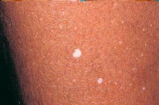
Clinical manifestations of hypomelanosis are characterized by the appearance of spots on the skin of white color and rounded shape. The diameter of such altered areas reaches up to 1 cm.
Drop-shaped hypomelanosis first appears on the shin (extensor surface), and then spread to the forearms, upper back and thorax. For this pathological condition is not characterized by damage to the skin of the face.
Progression of the process is observed with age, as well as with excessive exposure to direct sunlight.
Hito's Hypomelanoz
Pathology is observed in men and especially women and in terms of prevalence is second only to neurofibromatosis and tuberous sclerosis. Hypomelanosis is the result of sporadic diseases, but inheritance by recessive and dominant type is not excluded.
At the heart of the development of pathology is the failure of cell migration from the neural tube in the prenatal period, resulting in an abnormal location of gray matter in the brain, as well as an inadequate number of melanocytes in the thickness of the skin.
Migrating with melanoblast occurs in the second-third trimester of pregnancy. At the same time, the movement of neurons is observed, as a result of which the hypomelanosis of the otho includes clinical manifestations of pigmentation disorders and brain pathology.
Skin symptoms are expressed by areas of hypopigmentation of irregular shape (curls, zigzags, waves). Most of these foci are located near the Blaschko lines, and their appearance can be observed already in the first days or months of the baby's life, but before the adolescence they may become less noticeable or even disappear altogether.
Neurological symptoms are characterized by mental disorders, epileptic seizures, which are distinguished by their resistance to anticonvulsant therapy. Often kids suffer from autism, muscle hypotension and motor disinhibition. In a quarter of cases, macrocephaly is noted.
In addition, often a pathology of other organs is observed, for example, heart disease, anomaly of the structure of the genital organs, face, deformities of the spinal column, stop, eye symptoms, as well as disruption of the structure and growth of teeth and hair.
Idiopathic hypomelanosis
At the heart of the development of hypomelanosis is a violation of the stages of melanin synthesis due to the absence of melanocytes, a failure in the formation of full-fledged melanomas and their migration.
Melanocytes are derived from ectomesenchyma. Their differentiation goes through 4 stages. The first is the appearance of precursors of melanocytes in the neural scallop, the second is the migration of melanocytes in the thickness of the dermis towards the basal membrane of the epidermis. Further, their movement in the epidermis is noted, and, finally, the stage of formation of the processes (dendritic), when the cell occupies its positions in the epidermis.
Idiopathic hypomelanosis develops in the event of a breakdown in one of the listed stages, as a result of which melanocyte can be located in an unusual place for him, because of which a certain area of skin remains "colorless", since the pigment synthesis will be absent.
It can occur in toddlers or with age. In addition, when exposure to ultraviolet rays, the progression of this pathology is possible.
The main reason for the development of the disease is difficult to identify, since almost 100% of cases - it's a genetic breakdown. Idiopathic teardrop hypomelanosis can occur immediately after birth or during adolescence. Most often, the pathology has a chronic type of flow with periodic relapses.
Clinical manifestations of the disease are foci of hypopigmentation of different localization (shin, forearm, back) and a diameter of up to 1 cm. The sites are located separately from each other and are not capable of fusion.
Most often idiopathic guttate hypomelanosis is observed in women who have a light shade of skin, especially those living in an area with increased exposure to sunlight. In addition, at the first appearance of the focus on the shin, then under the influence of insolation of the sites of depigmentation becomes more and more.
Pathogenetic therapy aimed at removing the causative factor does not exist, and therefore symptomatic treatment is used to reduce the intensity of manifestations of pathology.
Diagnosis of hypomelanosis
Violation of the processes of pigmentation can manifest itself in various forms. For the verification of pathology, in addition to a visual examination, it is necessary to use research using the Wood lamp. Especially often it is used in the presence of light skin and indistinctly manifested pathology.
Diagnosis of hypomelanosis is based on the identification of clear boundaries of the hypopigmented focus in the process of glowing the lamp in a darkened room. Thanks to it, it becomes possible to locate the site and verify it.
Diagnosis of hypomelanosis Ito additionally includes a computerized tomography of the brain, where an increase in the 3rd and lateral ventricles, unclear boundaries between the brain substance, as well as atrophy of the frontal lobes.
Histological examination barks the possibility of revealing an inadequate amount of melanocytes on the hypopigmentation site. In addition, hypomelanoid Ito may have other features in the outbreak, for example, vascular nevuses, cocoa spots, nevus Ott or Mongoloid blue spots.
How to examine?
Who to contact?
Treatment of hypomelanosis
This pathological process is characterized by its spread at the genetic level, in connection with which pathogenetic treatment does not yet exist. Symptomatic therapy is used, the main directions of which are to stop the generalization of pathology and reduce its clinical manifestations.
Treatment of hypomelanosis involves the use of corticosteroids, the introduction of which is carried out inside the hearth. In addition, various studies have shown the effectiveness of the use of local retinoids, pimecrolimus (Elidel), as well as cryomassage of the affected areas.
Also, hypomelanosis can be treated with phototherapy, thanks to which activation of melanin production by pigment cells is noted. In addition, substitution therapy with melagenin is recommended. Its action is aimed at stimulating melanocytes to synthesize the pigment.
As for bioresonance therapy, it is aimed at restoring the normal functioning of the nervous system, as well as increasing the level of the body's immune forces.
Alternative treatment is also possible with this type of pathology, but before using it, consult a doctor.
Prevention of hypomelanosis
Specific prevention of hypomelanosis does not exist, since pathology has a genetic type of inheritance. However, there are still methods that can reduce the risk of developing hypomelanosis or the occurrence of its relapse.
The main provoking factor of generalization and progression of the process is considered excessive insolation. As a result, it is necessary to inform the population about its negative effect not only on hypomelanosis, but also on the likelihood of developing cancer.
Prevention of hypomelanosis consists in avoiding exposure to direct sunlight on unprotected skin, especially between 11.00 and 16.00, the use of sunscreen cosmetics in hot weather, since UV radiation can be reflected from surrounding objects and earth, passing through clouds and clothing. Because of this, it is not recommended to be on the street in the daytime in the absence of an acute need .. Also this applies to sunbeds in the solarium. To protect the skin, you need to use a special cream, hat and clothes that covered areas of hypomelanosis.
Prognosis of hypomelanosis
In themselves, areas of hypopigmentation in the form of white spots do not pose a health hazard, however, when the first clinical symptoms appear, you should consult a specialist for further diagnosis and treatment. The earlier the pathology is detected, the greater the probability of stopping the process and preventing the development of relapses.
The prognosis of hypomelanosis is favorable, but with excessive sunlight exposure, it can spread to healthy tissues, since excessive insolation helps reduce the amount of melanosomes and pigment.
It is impossible not to warn about the possibility of developing a carcinogenic process under the influence of sunlight. This is due to malignant cell degeneration due to violations of gene expression. In addition, each person has birthmarks that are also capable of changing under the influence of the sun.
So, hypomelanosis is not a terrible pathology, but still requires a special approach and compliance with specific measures to prevent the onset and relapse in chronic course.


 [
[