
Medical expert of the article

New publications
Vaginal atresia: causes, symptoms, diagnosis, treatment, and prognosis
Last updated: 24.03.2026

All iLive content is medically reviewed or fact checked to ensure as much factual accuracy as possible.
We have strict sourcing guidelines and only link to reputable media sites, academic research institutions and, whenever possible, medically peer reviewed studies. Note that the numbers in parentheses ([1], [2], etc.) are clickable links to these studies.
If you feel that any of our content is inaccurate, out-of-date, or otherwise questionable, please select it and press Ctrl + Enter.
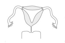
Vaginal atresia is a rare congenital anomaly of the female reproductive tract, in which the vaginal canal is partially or completely absent or replaced by fibrous tissue, impairing the normal flow of menstrual blood and proper anatomical patency. In modern literature, this condition is typically considered a consequence of impaired embryonic development of the Müllerian structures and the urogenital sinus. Importantly for clinical practice, the external genitalia may appear normal, and the uterus, fallopian tubes, and ovaries are often intact. [1]
The term is often confused with vaginal aplasia in Mayer-Rokitansky-Küster-Hauser syndrome, transverse vaginal septum, and imperforate hymen. This is not simply a linguistic issue. The prognosis, the extent of the examination, the choice of surgery, and the discussion of future menstrual and reproductive function depend on the correct distinction. The American College of Obstetricians and Gynecologists clearly emphasizes that examination of the external genitalia and proper visualization are critical for distinguishing between distal vaginal atresia, transverse septum, urogenital sinus, and imperforate hymen. [2]
The condition most often becomes noticeable during puberty, when a girl has normal development of secondary sexual characteristics but no menstruation and cyclical lower abdominal pain. If the obstruction is located distally, menstrual blood cannot exit and accumulates first in the vagina, which can then distend the cervix, uterine cavity, and even the fallopian tubes. Therefore, vaginal atresia is not only an anatomical anomaly but also a potential source of severe pain, endometriosis, inflammatory complications, and decreased fertility. [3]
Although the anomaly is rare, it cannot be considered isolated. A 2025 review estimated the incidence at approximately 1 in 10,000–15,000 female newborns, with the authors noting that the true incidence is likely underestimated due to late diagnosis and difficulty distinguishing it from other vaginal and cervical anomalies. This is why the modern approach emphasizes early recognition, high-quality imaging, and treatment in a specialized center, rather than empirical attempts to "discover" the obstruction without a full understanding of the anatomy. [4]
Code according to ICD-10 and ICD-11
The International Classification of Diseases, 10th revision, does not contain a separate code for "vaginal atresia." Congenital forms are typically classified under Q52.4, Other congenital malformations of the vagina, because this section contains congenital vaginal anomalies that do not have their own separate subcategory. This is an important clarification, since the N89.5, Vaginal stricture and atresia, clinical modifications refer to acquired non-inflammatory disorders and should not replace the congenital anomaly. [5]
The International Classification of Diseases, 11th revision, has refined the classification. A separate code, LB42.5 "Stricture or atresia of vagina," is provided for congenital atresia or stricture of the vagina, which is located in the section on congenital structural anomalies of the vagina. This reflects a more modern approach, in which various types of vaginal anomalies, including absence of vagina, septum, imperforate hymen, and atresia, are classified into separate categories. [6]
| Clinical situation | International Classification of Diseases, 10th revision | International Classification of Diseases, 11th revision | Comment |
|---|---|---|---|
| Congenital vaginal atresia | Q52.4 | LB42.5 | In the 10th revision there is no separate title; the general heading of congenital malformations of the vagina is used. |
| Congenital absence of the vagina | Q52.0 or clarification according to clinical formulation | LB42.0 | This is no longer atresia, but the absence of a vagina. |
| Imperforate hymen | Q52.3 | LB42.4 | Important differential diagnosis |
| Acquired vaginal stricture or atresia | does not apply to congenital codes Q52 | does not belong to the block of congenital anomalies | In clinical systems, other categories are used for the acquired variant. |
Source for the table: official version of the International Classification of Diseases, 10th revision of the World Health Organization and the headings of the International Classification of Diseases, 11th revision, version 2025-01. [7]
Epidemiology
Vaginal atresia is a rare congenital anomaly. A modern review from 2025 estimates its incidence at approximately 1 in 10,000–15,000 female births. The authors emphasize that this is an approximate, not a definitive, figure, as some cases are recognized only in adolescence, and others may be mistakenly attributed to other congenital anomalies of the vagina and cervix. [8]
In practice, the disease is more often diagnosed during puberty rather than in newborns. The typical scenario is normal breast development and hair growth in the absence of menarche, increasing cyclical pain, a feeling of pelvic pressure, or the appearance of a mass due to hematocolpos. A 2025 review indicates that in some countries, the median age of diagnosis is approximately 12 years, and late diagnosis remains a common problem. [9]
The distribution of anatomical variants is also uneven. In the reviewed series of 67 patients, 37.3% had lower vaginal atresia, 53.7% had complete atresia, 1.5% had upper vaginal atresia, and 7.5% had fornix atresia. These data cannot be automatically generalized to the entire population, but they clearly demonstrate that clinically, "vaginal atresia" is not a single anatomical scenario, but several different variants. [10]
An important epidemiological feature is the association with other anomalies. A 2025 review described associations with other Müllerian defects, urinary tract anomalies, scoliosis, anorectal defects, and cardiac defects. This is why, when vaginal atresia is confirmed, modern examinations are usually not limited to the pelvic organs. [11]
Table 2. What is known about the frequency and detection rate
| Indicator | What is known |
|---|---|
| Estimated frequency | 1 case per 10,000-15,000 newborn girls |
| Typical age of detection | More often puberty than the neonatal period |
| Reason for late diagnosis | Normal external genitalia, rare anomaly, confusion with other forms of obstruction |
| Frequent debut | Primary amenorrhea and cyclic pelvic pain |
| Important feature | Frequent association with other Müllerian and urinary anomalies |
Source for table: 2025 review of vaginal atresia and magnetic resonance imaging recommendations for congenital anomalies of the female genital tract. [12]
Reasons
The immediate cause of vaginal atresia is a disruption in the embryonic formation and canalization of the vaginal canal. In the classic model, the upper portions of the vagina are connected to the Müllerian structures, and the lower portions to the urogenital sinus, so a defect at one of these stages can lead to partial or complete obstruction. A modern review from 2025 emphasizes that congenital vaginal atresia is considered to be the result of abnormal development of the urogenital sinus and Müllerian ducts with incomplete canalization of the vagina. [13]
The cause is not always limited to a local vaginal anomaly. Complete atresia often involves a concomitant developmental disorder of the cervix, and sometimes a more complex uterine configuration. This is why, in some patients, the surgical goal is limited to restoring the opening of the normally formed uterus and upper vagina, while others require a much more complex reconstruction or even a reconsideration of the uterine preservation issue. [14]
The genetic basis is incompletely understood, but a 2025 review described associations with TBX6 variants, genes of the Tyro3 RTK family, LHFPL2, ADAMTS18, and some chromosomal rearrangements, including regions with HNF1B and LHX1. These data do not yet provide a simple clinical test for most patients, but they indicate that the anomaly, at least in some patients, has a genetic vulnerability. [15]
Environmental and maternal factors are discussed but are not yet considered as definitively proven as for many other congenital conditions. The same review notes that the influence of drugs, toxic factors, and maternal factors is possible, but the specific mechanisms and causal relationships remain insufficiently confirmed. Therefore, it is more accurate to speak not of "definitely known external causes," but of potential influences that are still being studied. [16]
Risk factors
There are no clearly established modifiable risk factors for vaginal atresia, such as those for diabetes or cardiovascular disease. Current data point more to a congenital embryological and, possibly, genetic basis for the anomaly. Therefore, in this context, the risk factor is often not the patient's behavior, but rather the underlying developmental characteristics of the fetus and the family. [17]
A clinically important factor of suspicion is a family history of congenital anomalies of the genital tract, kidneys, and urinary system. Although specific familial patterns have not been established for all cases, a 2025 review emphasizes the role of genetic susceptibility and chromosomal rearrangements in some patients. In everyday practice, this means that familial cases and associated defects require a more careful genetic and anatomical evaluation. [18]
The second practical risk factor is the presence of other congenital anomalies, primarily of the urinary system and the broader complex of Müllerian defects. European guidelines for magnetic resonance imaging emphasize that congenital anomalies of the female reproductive tract are often combined with anomalies of the kidneys and other organs, so identifying one anomaly increases the likelihood of finding another. [19]
Adolescents with primary amenorrhea and cyclic pain require special attention. This is not a "risk factor" in the strict epidemiological sense, but a clinical marker of a high probability of an obstructive anomaly, for which investigation should not be delayed. The American College of Obstetricians and Gynecologists (ACOG) recommends that if an obstructive anomaly is suspected, a history, examination, and imaging are necessary. In cases of distal vaginal atresia, the risk of stenosis and complications makes referral to an expert center particularly important. [20]
Table 3. Practical caution factors
| Factor | Why is it important? |
|---|---|
| Family history of congenital anomalies of the genital tract | Supports the innate and probably genetic nature of the process |
| Combined anomalies of the urinary system | Often found together with defects of the Müllerian structures |
| Primary amenorrhea with normal development of secondary sexual characteristics | Typical signal of obstructive anomaly |
| Cyclic pelvic pain without menstruation | Indicates possible menstrual obstruction |
| Previously detected abnormality of the uterus or cervix | Increases the likelihood of a complex combined anomaly |
Source for table: 2025 review, American College of Obstetricians and Gynecologists guidelines, and European Society of Urogenital Radiology guidelines.[21]
Pathogenesis
The pathogenesis of vaginal atresia is associated with the failure of the vaginal canal to form a complete lumen. Anatomically, this may manifest as a short, blind canal, a distal fibrous barrier, a long atretic segment, or a complete lack of normal patency between the uterus and the vaginal vestibule. Depending on the level of blockage, the clinical presentation and surgical approach will vary. [22]
If the patient has a functioning endometrium and an intact uterus, mechanical obstruction of the menstrual flow occurs after the onset of puberty. This initially leads to hematocolpos, followed by hematocervix, hematometra, hematosalpinx, and even hemoperitoneum. This is why the disease often manifests not at birth, but in adolescence, when the endometrium begins to function. [23]
Retrograde blood flow into the fallopian tubes and abdominal cavity has long-term consequences. A 2025 review emphasizes that endometriosis and pelvic adhesions can develop if detected late. In the cohort study cited in the review, endometriosis was detected in 66.7% of patients with distal vaginal hypoplasia and in 100% of patients with cervicovaginal hypoplasia, although these figures are specific to a specific series and should be interpreted with caution. [24]
If atresia is combined with a cervical anomaly, the pathogenesis becomes even more complex. In this case, the problem lies not only in the absence or closure of the vaginal lumen, but also in the disruption of the connection between the uterus and the lower genital tract. This is why, with complete atresia, especially with cervical dysgenesis, the prognosis for maintaining menstrual function and fertility is significantly worse than with the isolated distal form. [25]
Symptoms
The most typical manifestation in adolescence is primary amenorrhea with normal breast development and other secondary sexual characteristics. If the uterus is functional, this is almost always accompanied by cyclical lower abdominal pain, which recurs monthly and gradually becomes more severe. In some patients, a palpable or visualized pelvic mass associated with a blood collection is present. [26]
In distal atresia, the upper vagina, cervix, and uterine body may be normally formed, so blood quickly accumulates above the obstruction and symptoms appear early. A 2025 review emphasizes that such patients often present earlier due to more severe pain and a noticeable mass. This typically facilitates timely diagnosis and makes reconstruction less complex. [27]
With complete atresia, symptoms may progress more slowly, especially if there is a concurrent cervical anomaly. In this case, abdominal pain may be less specific, masses less noticeable, and seeking medical attention may be delayed. This type of atresia is often associated with more complex surgery, a higher incidence of endometriosis, and poorer reproductive outcomes. [28]
The partial form may present not only with amenorrhea but also with dysmenorrhea, difficulty with intercourse, prolonged or unusual menstrual flow, recurrent bleeding, or infections. Orphanet lists symptoms as primary amenorrhea, cyclic pelvic pain, abdominal pain, dyspareunia, pelvic mass, menstrual irregularities, and periodic fever. This is an important reminder that the clinical picture does not always fit the "no periods and severe pain" pattern. [29]
Rarely, the anomaly manifests itself in newborns or young children. According to a 2025 review, under the influence of maternal estrogens, some infants may experience distension of the uterine cavity and cervix, leading to obstructive urogenital symptoms, including urologic obstruction, urinary tract infection, and sepsis. This is a rare but important scenario that should not be overlooked when a pelvic cyst and urinary symptoms are combined in a newborn girl. [30]
Table 4. Typical symptoms by age and form
| Situation | What happens more often? |
|---|---|
| A teenager with a functioning uterus | Primary amenorrhea, cyclic pain, pelvic mass |
| Distal atresia | Earlier and more pronounced obstructive onset |
| Complete atresia with neck anomaly | Later diagnosis, more complex clinical scenario |
| Partial form | Dyspareunia, painful periods, outflow disorders |
| Neonatal period, rare variant | Urological obstruction, infection, pelvic cyst |
Source for table: 2025 survey and Orphanet database. [31]
Classification, forms and stages
There is no single, universal "staging" for vaginal atresia. In practice, it is more convenient to classify the disease by anatomical level, extent of obstruction, and association with cervical and uterine anomalies. A recent review from 2025 emphasizes that the distinction between distal atresia and complete atresia is particularly useful for clinical communication, as it best reflects the differences in surgical complexity and reproductive prognosis. [32]
In the system of the European Society of Human Reproduction and the European Society of Gynecological Endoscopy, vaginal atresia is classified under the additional vaginal class V4, while the anomaly of the uterus and cervix itself is coded separately. This is convenient for complex combined cases, when it is necessary not only to say "there is vaginal atresia," but to describe the entire architecture of the defect. Magnetic resonance imaging guidelines explicitly emphasize that a structured description of the uterus, cervix, vagina, ovaries, and kidneys facilitates accurate classification and surgical planning. [33]
The 2021 American classification system for Müllerian anomalies also expanded the description of vaginal and cervical anomalies compared to older schemes. The authors of the document emphasize that previous classifications focused too heavily on the uterus and inadequately addressed vaginal and cervical anomalies, leading to diagnostic errors, chronic pain, and loss of reproductive function. This directly applies to vaginal atresia. [34]
From a clinical perspective, it's especially important to differentiate between those with a functioning uterus and those without the prospect of uterine preservation. If the uterus and endometrium are intact, the goal of treatment is to restore the outflow and, if possible, future reproductive function. However, if there is a severe combined cervicovaginal anomaly, treatment becomes much more complex, and the decision to preserve the uterus requires careful consideration. [35]
Complications and consequences
The main early complications are associated with menstrual obstruction. These include hematocolpos, hematometra, hematosalpinx, hemoperitoneum, severe pain, and secondary inflammation. The longer the outflow obstruction persists, the higher the risk of retrograde menstruation and pelvic adhesions. Therefore, a wait-and-see approach without a clear plan is dangerous. [36]
One of the most serious long-term consequences is endometriosis. A 2025 review found that in one cohort series, endometriosis was detected in 66.7% of patients with distal vaginal hypoplasia and in 100% of patients with cervicovaginal hypoplasia. These figures cannot be mechanically generalized to all populations, but they clearly demonstrate the dangers of late relief of obstruction. [37]
After surgery, the main problem is restenosis, or re-closure of the vaginal canal. The American College of Obstetricians and Gynecologists clearly states that with distal vaginal atresia, transverse septum, and cervical atresia, the risk of stenosis and complications is high, so such patients should be treated in an experienced center. A recent review from 2025 adds that prolonged postoperative dilation using dilators or forms is necessary for at least several months, otherwise the risk of re-obstruction increases significantly. [38]
Reproductive outcomes depend on the type of anomaly. With the distal form, successful restoration of vaginal outflow usually leads to a significantly better prognosis. With complete atresia, and especially with associated cervical anomalies, pregnancy is not always possible. A 2025 review found that after surgery for cervicovaginal atresia, clinical pregnancy was achieved in 28.5% of patients, and live birth in 14%, highlighting the complexity of this group. [39]
The psychosocial consequences also cannot be underestimated. The same review reported that moderate to severe depressive symptoms were observed in 58.7% of patients with complete vaginal atresia, while anxiety, decreased self-esteem, and sexual adjustment problems persisted even after anatomically successful surgery. Therefore, modern care includes not only surgery but also psychological support. [40]
Table 5. Main complications and consequences
| Complication | Why does it arise? |
|---|---|
| Hematocolpos | Accumulation of blood above the level of obstruction |
| Hematometra and hematosalpinx | Continuation of obstruction with functioning endometrium |
| Endometriosis and pelvic adhesions | Retrograde menstruation and chronic inflammation |
| Restenosis after surgery | Scarring and re-closure of the canal |
| Infectious complications | Stagnation of content and complex reconstructions |
| Decreased fertility | Complex anatomy, endometriosis, cervical component |
| Psychological distress | Pain, amenorrhea, surgery, anxiety about sexual and reproductive function |
Source for table: 2025 review and recommendations of the American College of Obstetricians and Gynecologists.[41]
When to see a doctor
Evaluation should begin if a teenager has not menstruated by age 15 or if menstruation has not begun within 3 years of thelarche, especially if accompanied by cyclic pain, lower abdominal distension, a feeling of pelvic pressure, or a mass. Modern documents on amenorrhea consider these timeframes as the basis for assessing primary amenorrhea. In the case of vaginal atresia, delay is particularly undesirable, as the complaint of "painful periods without menstruation" may mask developing menstrual obstruction. [42]
Immediate in-person care is required for increasing pelvic pain, urinary retention, fever, vomiting, severe bloating, signs of infection, or a sudden deterioration in condition. Such symptoms may indicate massive hematocolpos, urologic compression, an inflammatory complication, or other consequences of obstruction. Although the American College of Obstetricians and Gynecologists notes that most obstructive anomalies are not immediate surgical emergencies, severe symptoms warrant prompt referral to a specialist. [43]
Once the diagnosis has been established, re-examination is necessary if pain recurrs, drainage decreases after reconstruction, dysmenorrhea develops, difficulty using dilators, signs of infection, or urination deteriorates. Recurrence of obstruction remains one of the main late problems, so postoperative follow-up should be proactive rather than pro forma. [44]
Diagnostics
Diagnosis begins with a history and a gentle but thorough examination. The American College of Obstetricians and Gynecologists emphasizes that a genital examination is critical for distinguishing between imperforate hymen, labial adhesions, urogenital sinus, transverse vaginal septum, and distal vaginal atresia. In practice, this means that blood tests or ultrasound alone are often insufficient without an examination. [45]
Pelvic ultrasound is usually the first line of imaging. It helps identify hematocolpos, hematometra, dilated fallopian tubes, large blood clots, and the overall configuration of the uterus. However, ultrasound has limitations: before puberty, the uterus is small, and complex combined vaginal and cervical anomalies may be incompletely or incorrectly described. This is specifically noted by both the American College of Obstetricians and Gynecologists and a 2025 review. [46]
If the anatomy is complex or precise preoperative mapping is required, magnetic resonance imaging (MRI) is the next step. European guidelines for imaging of congenital anomalies of the female genital tract (CAT) identify MRI as a method that allows for a comprehensive assessment of the uterus, cervix, vagina, kidneys, and complications in a single examination. A 2025 review explicitly calls MRI the "gold standard" for the assessment of complex Müllerian anomalies. [47]
Next comes the clarification of associated anomalies. In a structured magnetic resonance imaging (MRI) evaluation, it is recommended to assess not only the uterus, cervix, and vagina, but also the kidneys, ovaries, and complications. This is because vaginal anomalies are often associated with urinary tract anomalies. In complex cases and suspected endometriosis or other pelvic pathology, laparoscopy may be required, which is also reflected in the 2025 review. [48]
A karyotype, hormonal profile, and genetic evaluation are not required for everyone, but rather when the differential diagnosis includes Mayer-Rokitansky-Küster-Hauser syndrome, disorders of sexual development, or combined congenital syndromes. For typical distal atresia with a normal female phenotype and obvious obstruction, examination and imaging play a key role. However, in questionable cases, another anomaly cannot be overlooked. [49]
Table 6. Step-by-step diagnostic route
| Step | What is being assessed? | Why is this necessary? |
|---|---|---|
| 1 | Anamnesis | Understand the nature of pain, the presence of amenorrhea, cyclicity, urinary and intestinal symptoms |
| 2 | Examination of the external genitalia | Distinguish atresia from imperforate hymen, septum and other causes |
| 3 | Ultrasound examination of the pelvis | Find hematocolpos, hematometra, evaluate the uterus and tubes |
| 4 | Magnetic resonance imaging | Accurately map the level and extent of the anomaly, associated defects and complications |
| 5 | Evaluation of the kidneys and urinary tract | Exclude combined anomalies of the urinary system |
| 6 | Genetics and karyotype according to indications | Clarify complex and differential forms |
| 7 | Laparoscopy according to indications | Assess endometriosis, adhesions, and complex anatomy |
Source for table: American College of Obstetricians and Gynecologists guidelines, European Society of Urogenital Radiology guidelines, and 2025 review. [50]
Differential diagnosis
The most common differential diagnosis is an imperforate hymen. With this condition, the obstruction is located even more distally, and a bulging, bluish hymen may be visible during examination. The American College of Obstetricians and Gynecologists and a 2025 review emphasize that a thorough examination is most helpful here. With vaginal atresia, unlike with an imperforate hymen, the obstructive segment is usually located deeper, and a pronounced bluish bulge at the introitus may not be present. [51]
The second important variant is a transverse vaginal septum. Clinically, it can also cause primary amenorrhea and cyclic pain, but anatomically, it is not atresia, but the presence of a septum between normally formed sections of the vagina. This is a very important distinction for surgery: with a septum, the surgical approach is different, and the risk of stenosis remains high. Therefore, both conditions should not be confused under the common term "vaginal closure." [52]
The third major differential diagnosis is Mayer-Rokitansky-Küster-Hauser syndrome, which is aplasia or agenesis of the vagina with the absence or severe underdevelopment of the uterus. A 2025 review emphasizes that this syndrome typically lacks hematocolpos and does not present with significant menstrual obstruction, as a functioning uterus is often absent. This is why, in such patients, primary amenorrhea is associated not so much with cyclic obstructive pain as with the anatomical inability to menstruate. [53]
Finally, we must not forget about more complex combined anomalies, including cervical atresia, obstructive hemi-vaginal syndrome with ipsilateral renal anomaly, and other Müllerian defects. In complex cases, the only reliable approach is not to guess the diagnosis by appearance, but to base it on a combination of examination, ultrasound, and magnetic resonance imaging with the participation of an experienced radiologist and surgeon. [54]
Treatment
Treatment of vaginal atresia is surgical in most cases. A modern review from 2025 and a prospective randomized trial from 2025 clearly indicate that in congenital vaginal atresia, surgery is an effective way to eliminate anatomical obstruction. Unlike isolated vaginal agenesis in Mayer-Rokitansky-Küster-Hauser syndrome, where dilation therapy is often the primary option, in true atresia with menstrual obstruction, restoration of patency is usually required. [55]
The first principle of treatment is to delay technical intervention until the anatomy is fully understood. The American College of Obstetricians and Gynecologists emphasizes that the best long-term outcome is achieved with a complete examination, a clear understanding of the anomaly, proper preoperative preparation, and elective surgery at a center experienced in treating such defects. This is especially important due to the high risk of stenosis and restenosis in distal vaginal atresia, transverse septum, and cervical atresia. [56]
In cases of distal vaginal atresia, the treatment goal typically involves dissecting the atretic segment, evacuating accumulated blood, and restoring a continuous channel between the superior vagina and the vestibule. According to review data, this option often yields the most favorable anatomical and reproductive outcomes, as the uterus, cervix, and superior vagina in such patients are often normally formed. However, even in this case, postoperative stenosis should not be underestimated. [57]
In cases of complete vaginal atresia, especially in combination with cervical dysgenesis, surgery becomes significantly more complex. Vaginoplasty, cervicovaginal anastomosis, or uterovaginal anastomosis are already being considered in carefully selected cases, provided there is a well-developed uterus and the patient is interested in preserving menstrual and reproductive function. However, a 2025 review emphasizes that it is precisely in this group that the rate of re-obstruction, complications, and unsatisfactory reproductive outcomes remains highest. [58]
A review of 33 studies of cervicovaginal anastomosis in 53 patients showed menstrual return in 62.3% and only 1 natural pregnancy. Among 121 patients undergoing uterovaginal anastomosis, menstrual return was 77.7%, but serious complications also occurred, including fistula and subsequent hysterectomy in some patients. These figures demonstrate that uterine-preserving surgery is possible, but requires extremely careful selection and honest communication about the risks. [59]
Reconstructive techniques described include peritoneal vaginoplasty, intestinal vaginoplasty, amniotic grafting, robotic approaches, and biomaterial techniques. A 2025 review described peritoneal vaginoplasty as an effective and relatively cost-effective technique with high satisfaction, while intestinal vaginoplasty was described as an option with good patency and a low risk of stenosis, but a more invasive one. All this demonstrates that "one best procedure for all" does not exist. The method is selected based on the type of anomaly, age, length of the atretic segment, condition of the cervix, center experience, and reproductive goals. [60]
New approaches include laparoscopic reconstruction with peritoneal flaps and cervicoplasty, as well as modified vaginal repair techniques. In pilot and early clinical trials in 2024 and in a prospective randomized trial in 2025, some new techniques have shown fewer stenosis, shorter operative times, and better satisfaction rates, but these data are still primarily from individual centers and require broader independent validation. Therefore, such methods can be considered promising, but not as an absolute universal standard. [61]
Postoperative vaginal dilation is a critical part of treatment, not an afterthought. A 2025 review emphasizes that with any reconstructive technique, regular use of a vaginal dilator or form for at least 6 months is crucial to prevent restenosis and re-obstruction. The same review also reports that patients who used dilators regularly had longer vaginal canal lengths and better sexual outcomes than those who used them infrequently. [62]
Psychological support is equally important. In a review of the literature, moderate or severe depressive symptoms were reported in 58.7% of patients with complete vaginal atresia. Furthermore, surgery, dilators, anxiety about future sexual and reproductive function, and family stress can all impair treatment adherence. Therefore, a modern care pathway should include not only a surgeon and radiologist, but also a specialist in adolescent gynecology and, if necessary, a psychologist or psychotherapist. [63]
In terms of fertility, the prognosis depends heavily on the type of anomaly. With distal atresia, natural pregnancy often remains possible after adequate vaginal reconstruction. With complete atresia with a cervical component, the outcomes are much worse: a 2025 review cites a clinical pregnancy rate of 28.5% after reconstruction and a live birth rate of 14% in patients with cervicovaginal atresia. Therefore, when discussing treatment, it is important to immediately distinguish between two goals: creating a functional vagina and attempting to preserve the uterus and reproductive potential. This is not always achieved with the same surgery and is not always possible with equal success. [64]
Table 7. Main approaches to treatment
| Approach | Where is it most often used? | Key Features |
|---|---|---|
| Dissection and restoration of the distal canal | Distal atresia | Usually the best functional prognosis |
| Cervicovaginal anastomosis | Combined cervicovaginal anomaly | Possibility of maintaining outflow, but high risk of restenosis |
| Uterovaginal anastomosis | Individual complex cases | Menstrual flow is possible, but there are more complications |
| Peritoneal vaginoplasty | Complete atresia and complex reconstructions | An effective reconstructive option, including laparoscopic |
| Intestinal vaginoplasty | Selected cases | More invasive but with a low risk of stenosis |
| Amniotic and other biomaterials | Specialized centers | Promising, but depends on experience and availability |
| Postoperative dilators or forms | After almost any reconstruction | Needed for the prevention of stenosis |
| Psychological help | All patients with significant stress and complex treatment | Improves adherence and quality of life |
Source for table: 2025 review, ACOG 2019, and 2025 randomized trial. [65]
Prevention
There is currently no specific primary prevention for congenital vaginal atresia, as it is an embryological anomaly rather than a lifestyle disease. Recent reviews emphasize that the genetic and embryonic basis is incompletely understood, and no convincingly proven modifiable factors have been identified in most patients. Therefore, modern medicine cannot yet guarantee the prevention of this anomaly. [66]
However, secondary prevention of complications is entirely possible. This involves early recognition of primary amenorrhea, timely imaging, referral to an experienced center, careful selection of the initial surgery, and mandatory postoperative dilation of the urethra. This approach best reduces the risk of endometriosis, pelvic adhesions, infections, and recurrent obstruction. [67]
Prevention of adverse outcomes also includes a comprehensive search for associated anomalies, including those of the kidneys and urinary tract, and early psychological support. In a modern interdisciplinary approach, prevention is no longer an attempt to "prevent a defect," but rather to avoid its delayed recognition and severe consequences. [68]
Forecast
The prognosis depends primarily on the anatomical type of the anomaly. With distal vaginal atresia, if the diagnosis is made before severe endometriosis and the surgery is performed in a specialized center, the prognosis is usually more favorable: menstrual flow can be restored, a functional channel can be created, and a good chance of sexual and often reproductive function can be maintained. A 2025 review directly contrasts the more favorable outcome with the distal form with the significantly more complex prognosis with complete atresia. [69]
In cases of complete atresia, especially if there is a cervical anomaly, the prognosis is significantly more cautious. Even if anatomical reconstruction is successful, the risk of recurrent obstruction, infectious complications, and secondary uterine loss remains significant, and pregnancy is not possible for everyone. Therefore, realistic counseling and long-term follow-up are essential for this group. [70]
Long-term functional outcome depends not only on the surgery but also on postoperative management, particularly the use of dilators. A 2025 review emphasizes that regular postoperative dilation is associated with improved canal length and more satisfactory sexual outcomes. This suggests that prognosis is partly determined not only by anatomy but also by the quality of subsequent care. [71]
The psychological prognosis also cannot be automatically considered favorable after a successful operation. Current data show that even with improved sexual function, anxiety, depressive symptoms, and self-esteem issues may persist. Therefore, a good outcome for vaginal atresia is now understood more broadly than simply "an open vagina": it includes menstrual function, pain relief, stable patency, sexual adaptation, psychological well-being, and, if possible, preservation of reproductive prospects. [72]
FAQ
Are vaginal atresia and vaginal aplasia the same thing?
No. Atresia typically refers to a partial or complete closure or non-canalized segment of the vagina, often in the presence of the uterus and sometimes the upper vagina. Vaginal aplasia or agenesis is more commonly associated with Mayer-Rokitansky-Küster-Hauser syndrome, where the vagina and often the uterus are absent or severely underdeveloped. [73]
Why does a girl have breasts but not menstruation?
Because the ovaries and hormonal development are often preserved in vaginal atresia, and the problem lies in anatomical obstruction of the outflow. Therefore, secondary sexual characteristics develop normally, but menstrual blood cannot escape. [74]
Is this always discovered only in adolescence?
Most often, yes, but not always. In rare cases, the anomaly is noticed in newborns due to a pelvic cyst, urological obstruction, or infection, especially under the influence of maternal estrogens. [75]
Is ultrasound alone enough?
Not always. Ultrasound is usually the first step, but in complex anatomy, magnetic resonance imaging provides much more accurate mapping of the vagina, cervix, uterus, and associated anomalies. [76]
Is it possible to avoid surgery?
In true vaginal atresia, menstrual obstruction is usually absent. In such cases, surgical restoration of patency is required. Non-surgical dilation is the standard treatment for vaginal agenesis without obstruction rather than for classic atresia. [77]
How often does the stenosis re-close after surgery?
The risk of restenosis is considered a major one. This is why ACOG recommends treatment in an experienced center, and a 2025 review emphasizes the importance of regular use of dilators or forms after surgery. [78]
Is it possible to become pregnant later?
This depends on the type of anomaly. With distal atresia, the prognosis is usually better. With complete atresia with cervical involvement, the results are significantly worse. According to review data for cervicovaginal atresia after reconstruction, clinical pregnancy was achieved in 28.5% of patients, and live birth in 14%. [79]
Why is psychological help so important?
Because the disease affects menstrual function, body image, sexuality, and fertility concerns, and often requires long-term treatment with dilators and follow-up examinations. Recent reviews show a high rate of depressive symptoms and the need for psychological support before and after surgery. [80]
Key points from experts
Samantha M. Pfeifer, MD, professor of clinical obstetrics, gynecology, and clinical reproductive medicine at Weill Cornell Medical College, is an expert on Müllerian anomalies. Her expertise is particularly important for the topic of classification. The key practical thesis is this: a more accurate and comprehensive classification system is needed for congenital anomalies of the genital tract, because older, simplified classification schemes were less accurate in accounting for vaginal and cervical defects, thereby contributing to erroneous surgeries, chronic pain, and loss of reproductive function. [81]
Marc R. Laufer, MD, chief of gynecology at Boston Children's Hospital, co-director of the Center for Young Women's Health, professor at Harvard Medical School, and a renowned expert in congenital anomalies of the reproductive tract. His clinical thesis is particularly important for adolescent practice: congenital anomalies of the vagina and uterus should be recognized early and managed in a center with expertise in adolescent gynecology, endometriosis, and reconstructive surgery. For vaginal atresia, this means that an isolated "minor" examination without a comprehensive workup is often insufficient. [82]
Cristina Maciel, MD, Professor of Urogenital Imaging at the Faculty of Medicine, University of Porto, and member of the Female Pelvic Imaging Working Group of the European Society of Urogenital Radiology. Her expert opinion is reflected in the European Society of Urogenital Radiology guidelines: magnetic resonance imaging should not simply “confirm anomalies” but should fully map the uterus, cervix, vagina, kidneys, and complications within a standardized protocol and structured reporting. This makes preoperative planning safer and more accurate. [83]
Conclusion
Vaginal atresia is a rare but clinically significant congenital anomaly that most often presents with primary amenorrhea and cyclic pelvic pain in the context of normal pubertal development. It should not be confused with imperforate hymen, transverse vaginal septum, and Mayer-Rokitansky-Küster-Hauser syndrome, as these conditions have different anatomy, different treatments, and different prognoses. The best outcome is achieved when the diagnosis is made before the development of severe endometriosis and adhesions, and surgery is planned after complete anatomical mapping. [84]
The modern care strategy is built around four pillars. These include early recognition of primary amenorrhea and cyclic pain, high-quality imaging with magnetic resonance imaging for complex cases, surgery in an experienced center, and mandatory long-term postoperative care with canal dilation and psychological support. With the distal form, the prognosis is usually significantly better, and with complete atresia with a cervical component, treatment and counseling must be especially careful and transparent. [85]