Medical expert of the article
New publications
Treatment of acute and chronic iridocyclitis
Last reviewed: 04.07.2025

All iLive content is medically reviewed or fact checked to ensure as much factual accuracy as possible.
We have strict sourcing guidelines and only link to reputable media sites, academic research institutions and, whenever possible, medically peer reviewed studies. Note that the numbers in parentheses ([1], [2], etc.) are clickable links to these studies.
If you feel that any of our content is inaccurate, out-of-date, or otherwise questionable, please select it and press Ctrl + Enter.

Depending on the cause of the inflammatory process of iridocyclitis, general and local treatment of iridocyclitis is carried out.
At the first examination of the patient it is not always possible to determine the cause of iridocyclitis. The etiology of the process can be established in the following days, and sometimes it remains unknown, but the patient needs emergency care: a delay in prescribing treatment even for 1-2 hours can seriously complicate the situation. The anterior and posterior chambers of the eye have a small volume, and 1-2 drops of exudate or pus can fill them, paralyze the exchange of fluid in the eye, glue the pupil and lens.
First aid
In case of inflammation of the iris and ciliary body of any nature, first aid is aimed at maximum dilation of the pupil, which allows solving several problems at once. Firstly, when the pupil dilates, the vessels of the iris are compressed, therefore, the formation of exudate decreases and accommodation is simultaneously paralyzed, the pupil becomes motionless, thereby providing rest to the affected organ. Secondly, the pupil is diverted from the most convex central part of the lens, which prevents the formation of posterior synechiae and provides the possibility of rupture of existing adhesions. Thirdly, a wide pupil opens an outlet into the anterior chamber for the exudate accumulated in the posterior chamber, thereby preventing the gluing of the ciliary body processes, as well as the spread of exudate into the posterior segment of the eye.
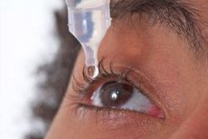
To dilate the pupil, instill 1% atropine sulfate solution 3-6 times a day. In case of inflammation, the duration of action of mydriatics is many times shorter than in a healthy eye. If synechia is already detected during the first examination, other mydriatics are added to atropine, for example, a 1:1000 adrenaline solution, a mydriacyl solution. To enhance the effect, a narrow strip of cotton wool soaked in mydriatics is placed behind the eyelid. In some cases, a crystal of dry atropine can be placed behind the eyelid. Non-steroidal anti-inflammatory drugs in the form of drops (naklof, diklof, indomethacin) enhance the effect of mydriatics. The number of combined mydriatics and instillations in each specific case is determined individually.
The next first aid measure is a subconjunctival injection of steroid drugs (0.5 ml dexamethasone). In case of purulent inflammation, a broad-spectrum antibiotic is administered under the conjunctiva and intramuscularly. To eliminate pain, analgesics and pterygopalatine-orbital novocaine blockades are prescribed.
Treatment regimen for iridocyclitis
Treatment of iridocyclitis depends on its cause, severity, and associated conditions. In general, therapy may include the following components:
Drug treatment:
- Topical corticosteroids (eg, prednisolone, dexamethasone) to reduce inflammation.
- Mydriatics (eg, atropine, cyclopentolate) to prevent adhesion formation and relieve pain by stabilizing the iris.
- Antibiotics or antiviral drugs in case of infectious etiology.
- Immunosuppressants and immunomodulators if an autoimmune process is confirmed.
Systemic treatment:
- Oral corticosteroids in cases of severe or resistant iridocyclitis.
- Immunosuppressive therapy (eg, methotrexate, azathioprine) to manage systemic inflammation, especially in associated autoimmune diseases.
Treatment of the underlying disease: If iridocyclitis is a manifestation of a systemic disease such as rheumatoid arthritis, Behcet's disease, or sarcoidosis, attention should also be directed to treating the underlying condition.
Monitoring and supportive therapy:
- Regular observation by an ophthalmologist to monitor the effectiveness of treatment and timely correction of therapy.
- Maintenance treatment aimed at reducing the risk of relapse.
Surgical treatment:
- In rare cases, if complications develop (such as cataracts or glaucoma), surgery may be required.
Patients with iridocyclitis should be monitored regularly by an ophthalmologist to adapt the treatment regimen according to their individual response to therapy and changes in the disease state.
Important: Before starting any therapy, it is necessary to undergo a complete medical examination and receive an accurate diagnosis. All treatment prescriptions must be made by qualified medical professionals.
Anticholinergics
Anticholinergics such as atropine and its derivatives (eg, scopolamine and homatropine) and synthetic drugs including cyclopentolate and tropicamide may be used to treat iridocyclitis. These medications act as mydriatics, causing pupil dilation, which helps with the following:
- Prevention of adhesions of the iris (posterior synechiae) with the lens, preventing their formation or resolving already formed adhesions.
- Relieves pain by stabilizing the iris and reducing pressure inside the eye.
- Reduce inflammation by stabilizing ocular tissues and preventing additional release of inflammatory mediators.
- Improving the drainage of fluid within the eye, which can help manage intraocular pressure.
It is important to note that the use of anticholinergics should be strictly under the supervision of an ophthalmologist, as they can cause side effects such as increased intraocular pressure (especially in patients with a narrow anterior chamber angle), blurred vision, photophobia and rarely systemic effects due to absorption through the conjunctiva.
In case of iridocyclitis, the dosage and duration of anticholinergic use will depend on the severity and progression of the disease.
Mydriatics
Mydriatics are drugs that cause pupil dilation and are often used in the treatment of iridocyclitis. Their use in iridocyclitis is necessary for several purposes:
- Preventing or breaking up adhesions between the iris and lens, known as synechiae, which may help avoid the development of secondary glaucoma or cataracts.
- Reduction of pain and discomfort caused by spasm of the iris muscles.
- Improved management of inflammatory exudate from the pupillary area, helping to reduce the risk of adhesions.
Classic mydriatics used in iridocyclitis include:
- Atropine: One of the most powerful mydriatics, also has a long-lasting effect. It is used for prolonged pupil dilation.
- Scopolamine: It has similar effects to atropine, but is less popular due to potential side effects.
- Cyclopentolate: A fast-acting mydriatic, usually used for short-term pupil dilation.
- Tropicamide: Another fast-acting mydriatic, it is usually used for diagnostic purposes and for short-term treatment of inflammatory eye diseases.
These drugs can be used in different concentrations and at different frequencies depending on the individual case and the recommendations of the attending physician. It is always necessary to carry out therapy under strict medical supervision, since mydriatics can increase the risk of developing an acute attack of glaucoma, especially in patients with a narrow angle of the anterior chamber of the eye.
Antibiotics
Antibiotics for iridocyclitis may be prescribed in cases where the inflammation is caused by bacteria or when there is a high risk of bacterial infection. The choice of a specific antibiotic depends on the suspected pathogen and its sensitivity to drugs.
Examples of antibiotics that may be used for bacterial iridocyclitis include:
Topical antibiotics (eye drops):
- Fluoroquinolones (eg, ofloxacin, levofloxacin)
- Aminoglycosides (eg, tobramycin, gentamicin)
- Macrolides (eg, erythromycin)
Oral antibiotics:
- Doxycycline or minocycline for infections caused by chlamydia or mycoplasma
- Cephalosporins or penicillins to combat a wide range of bacterial infections
Intravenous antibiotics:
- In cases of severe infections that are not controlled by topical or oral medications, stronger antibiotics such as vancomycin or ceftriaxone may be prescribed.
When treating iridocyclitis, it is very important to accurately determine the cause of the inflammation, since antibiotics are effective only against bacterial infections and are useless against viral, fungal, allergic or autoimmune processes. In some cases, laboratory tests may be required to determine the pathogen, including cultures from the mucous membrane of the eye and blood tests.
Antibiotic treatment should always be carried out under the supervision of an ophthalmologist and/or a physician. Incorrect use of antibiotics can lead to deterioration of the condition, development of resistance of microorganisms and other side effects.
Treatment of iridocyclitis in Bechterew's disease
Iridocyclitis associated with Bechterew's disease (ankylosing spondylitis) is an important ophthalmologic problem, as it can lead to serious visual impairment. It is an inflammation of the iris and ciliary body of the eye, which requires timely and adequate treatment. The approach to therapy is usually multidisciplinary and includes the following aspects:
Local treatment:
- Mydriatics (pupil dilators), such as atropine or cyclopentolate, to keep the pupil still and prevent the formation of posterior synechiae (adhesions) that may occur due to inflammation.
- Topical corticosteroids (such as prednisone) to reduce inflammation in the eye.
Systemic treatment:
- Nonsteroidal anti-inflammatory drugs (NSAIDs) to control the general inflammatory process in ankylosing spondylitis.
- Immunosuppressive drugs (eg, methotrexate) for more severe cases of both conditions.
- Biologic agents (TNF-alpha antagonists) such as infliximab or adalimumab, which have been shown to be effective in the treatment of both ankylosing spondylitis and related uveitis.
Control of the underlying disease:
- Managing the symptoms of ankylosing spondylitis may also help reduce the incidence and severity of iridocyclitis.
Monitoring and support:
- Regular follow-up with an ophthalmologist to assess the response to treatment and early detection of possible complications.
- Optimizing overall inflammation with physical therapy and exercises as recommended for ankylosing spondylitis may indirectly help improve iridocyclitis.
It is important to remember that the selection of drugs should be carried out individually, depending on the severity of the inflammatory process, the general condition of the patient and the presence of concomitant diseases. In addition, close contact between the patient, rheumatologist and ophthalmologist is important to achieve the best treatment results.
Treatment of herpetic iridocyclitis
Herpetic iridocyclitis is an inflammation of the anterior segment of the eye caused by infection with the herpes simplex virus (HSV) or the varicella-zoster virus (VZV). Treatment of this condition should be comprehensive and usually includes the following components:
Antiviral drugs:
- Oral antiviral drugs such as acyclovir, valacyclovir or famciclovir are the mainstay of therapy. They help reduce viral replication and limit its spread.
- Topical antiviral medications such as trifluridine or ganciclovir eye drops may also be used in some cases.
- In some severe or recurrent cases, antiviral drugs may need to be injected directly into the eye (periocular injections).
Anti-inflammatory drugs:
- Steroid eye drops (such as prednisolone) are used to reduce inflammation and prevent scarring.
- Caution: Steroids should be used with caution as they may enhance viral replication. Therefore, their use should be strictly supervised by an ophthalmologist.
Mydriatics (pupil dilators):
- To prevent the formation of posterior synechiae and to reduce pain and spasm of the ciliary body, mydriatic and cycloplegic agents such as atropine or cyclopentolate are used.
Supportive therapy:
- Use of artificial tears to reduce symptoms of dry eye caused by mydriatics or as a result of inflammation.
Monitoring and prevention of relapses:
- Regular eye examinations are important to monitor eye health and prevent chronic inflammation and relapses.
- Long-term preventive antiviral therapy may be recommended in cases of frequent relapses.
Treatment of concomitant complications:
- Such complications may include secondary glaucoma and cataracts, which may require specific medical or surgical treatments.
Treatment of herpetic iridocyclitis should be individualized and depends on the degree of inflammation, the presence of complications, and the patient’s overall health. It is important to begin treatment as soon as possible to reduce the risk of long-term vision problems.
Treatment of acute iridocyclitis
After the etiology of iridocyclitis is clarified, the identified foci of infection are sanitized, a general treatment plan is developed, prescribing agents that affect the source of infection or toxic-allergic influence. Correction of the immune status is carried out. Analgesics and antihistamines are used as needed.
In local treatment of iridocyclitis, daily correction of the therapy is necessary depending on the reaction of the eye. If it is not possible to rupture the posterior synechiae with the help of conventional instillations, then enzyme therapy (trypsin, lidase, lekozyme) is additionally prescribed in the form of parabulbar, subconjunctival injections or electrophoresis. It is possible to use medicinal leeches in the temporal region on the side of the affected eye. A pronounced analgesic and anti-inflammatory effect is provided by a course of pterygopalatine-orbital blockades with steroid, enzyme preparations and analgesics.
In case of a profuse exudative reaction, posterior synechiae may form even with pupil dilation. In this case, it is necessary to promptly cancel mydriatics and prescribe miotics for a short time. As soon as the adhesions have broken off and the pupil has narrowed, mydriatics are prescribed again ("pupil gymnastics"). After achieving sufficient mydriasis (6-7 mm) and synechiae have ruptured, atropine is replaced with short-acting mydriatics that do not increase intraocular pressure with prolonged use and do not cause side effects (dry mouth, psychotic reactions in the elderly). In order to exclude the side effects of the drug on the patient's body, it is advisable to press the area of the lower lacrimal point and lacrimal sac with a finger for 1 minute when instilling atropine, when the drug does not penetrate through the lacrimal ducts into the nasopharynx and gastrointestinal tract.
At the stage of calming the eye, magnetic therapy, helium-neon laser, electro- and phonophoresis with drugs can be used for faster resorption of the remaining exudate and adhesions.
Treatment of chronic iridocyclitis
Treatment of chronic iridocyclitis is long-term. The tactics of specific etiologic therapy and general strengthening treatment are developed jointly with a therapist or phthisiatrician. Local measures for tuberculous iridocyclitis are carried out in the same way as for diseases of other etiologies. They are aimed at eliminating the source of inflammation, resorption of exudate and preventing pupil overgrowth. With complete fusion and overgrowth of the pupil, they first try to break the adhesions using conservative means (mydriatics and physiotherapeutic effects). If this does not give a result, then the adhesions are separated surgically. In order to restore communication between the anterior and posterior chambers of the eye, laser pulsed radiation is used, with the help of which a hole (coloboma) is made in the iris. Laser iridectomy is usually performed in the upper root zone, since this part of the iris is covered by the eyelid and the newly formed hole will not give excess light.
References
Books:
- "Uveitis: Fundamentals and Clinical Practice" by Robert B. Nussenblatt and Scott M. Whitcup, 2010 edition.
- "Clinical Ophthalmology: A Systematic Approach" by Jack J. Kanski, 8th edition, 2016.
- "Ophthalmology" by Myron Yanoff and Jay S. Duker, 5th edition, 2018.
- "The Massachusetts Eye and Ear Infirmary Illustrated Manual of Ophthalmology" by Neil J. Friedman, Peter K. Kaiser, and Roberto Pineda II, 4th edition, 2014.
Research:
- "Treatment of Chronic Uveitis by Interferon-alpha" – authors Kramer M. and Pivetti-Pezzi P., published in "Ophthalmologica", 2000.
- "Efficacy and Safety of Immunosuppressive Agents in the Treatment of Noninfectious Intermediate, Posterior, and Panuveitis: A Systematic Literature Review" by Jabs DA, Nussenblatt RB, and Rosenbaum JT, published in the American Journal of Ophthalmology, 2010.
- "Anti-TNF Therapy in the Management of Acute and Chronic Uveitis" by Sfikakis PP, Theodossiadis PG, and Katsiari CG, published in Cytokine, 2002.
- "Biologic Therapies for Autoimmune Uveitis" by Pasadhika S. and Rosenbaum JT, published in "Ocular Immunology and Inflammation", 2014.


 [
[