
Medical expert of the article

New publications
Iridodiagnosis
Last reviewed: 03.07.2025

All iLive content is medically reviewed or fact checked to ensure as much factual accuracy as possible.
We have strict sourcing guidelines and only link to reputable media sites, academic research institutions and, whenever possible, medically peer reviewed studies. Note that the numbers in parentheses ([1], [2], etc.) are clickable links to these studies.
If you feel that any of our content is inaccurate, out-of-date, or otherwise questionable, please select it and press Ctrl + Enter.
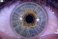
Indications for iridology
There are special indications for iridology. Thus, a number of changes in the human body can be noticed by the iris. Naturally, it is not so easy for an inexperienced person in this matter. But thanks to modern equipment and the qualifications of doctors, it is not so difficult.
Each part of the iris is responsible for its own organ or system. Therefore, it is easiest to see and determine diseases of the gastrointestinal tract, diseases of the spine, kidneys, genitals and cardiovascular system.
People with such unconfirmed diagnoses go to an experienced iridologist for help. It will not be difficult for the doctor to see all the changes in the body. In addition, you can see gastritis, ulcers, pancreatitis and other diseases.
It is amazing how the iris of the eye can tell about all this. That is why everyone who wants to uses the services of iridology. Because it is possible to see many diseases. This is what iridology is famous for, because it can do what traditional medicine cannot.
Preparation for iridology
Is preparation required for iridology, and what is needed for this? Unlike standard diagnostic methods for many diseases, this one does not require anything. A person just needs to make an appointment and visit it.
Naturally, we can talk about many diseases. Thus, during the determination of problems with the gastrointestinal tract, a number of measures are carried out. It is necessary to do an ultrasound of the abdominal cavity, for this a special solution is drunk, etc. There are also many procedures with the genitals.
As for iridology, no preparation is needed. A person simply comes, and an experienced doctor examines his eye with special equipment. That is the whole procedure. Therefore, there is simply no point in preparing, and there is no need for it.
The doctor examines the patient using various magnifying devices and examines the iris. Based on the data obtained, a diagnosis is made. In principle, that's all. So, iridodiagnosis is perhaps one of the simplest and most interesting methods of diagnosis.
Equipment for iridology
What equipment is needed for iridology? Naturally, special "devices" are purchased for this method. In fact, there are quite a few of them.
The essence of this equipment is that it allows you to see the iris of the eye in a certain color range. After all, this or that disease is determined precisely by the coloring in the areas of the eye. Modern equipment allows you to enlarge the iris, so that it is easier to understand what shades are inherent in this or that part of the eye.
In addition to the equipment itself, the diagrams also play an important role. They indicate which shade is responsible for what and which part of the iris indicates the presence of a particular disease.
Thanks to special devices, you can thoroughly examine the iris, take a picture of it for comparison and determine a diagnosis. The fact is that iridology is very young and has not yet earned universal recognition, but it is all a matter of time. After all, there is equipment, there are specialists, the method works.
 [ 3 ]
[ 3 ]
How is iridology performed?
Many patients are interested in how iridology is performed and what it is. So, an iridologist examines the iris of the eye using various magnifying devices. After that, he analyzes the obtained structure of the shell and compares it with special iridological schemes.
Based on the data obtained, the affected area of a specific organ is determined. But the most interesting thing is that the obtained schemes can also be used to determine a person's genetic predisposition to certain diseases.
As a rule, the doctor reads information from the eye for 7-10 minutes. The procedure is quite fast, and its efficiency is 95-100%. After the examination, the doctor sends the person with the diagnosis to a specific specialist. The latter, in turn, carries out his procedures and accurately determines what disease the patient has. In addition, he gives advice on how to get rid of it, prescribes medications and explains preventive measures. This is how interesting iridology is.
Computerized iridology
The method of determining diseases by the iris of the eye is called computer iridodiagnosis. Thanks to this procedure, it is easy to identify problems with the internal organs of a person, even at the initial stages. It is not always possible to do this using traditional methods.
In addition, it is possible to detect changes in organs, see the localization of the problem and prescribe timely treatment. Computerized iridology allows for decoding even in the most complex clinical cases.
For each patient, the genetic status and predisposition to hereditary diseases are determined individually. Computerized iridology combines all modern technological achievements. Anyone can be examined in the office of an experienced iridologist.
The data obtained can not only prevent the development of the disease, but also prescribe preventive measures. This is very convenient. The method is new, but over time it can become one of the most popular. Because iridology can cope with even severe cases.
[ 4 ]
Contraindications to iridology
Everyone can experience all the delights of the new method, because there are no contraindications to iridology. Indeed, you are free to come to the doctor's appointment. No need for any records, indications, etc.
After all, iridology can see what others cannot. It not only helps to make an accurate diagnosis for a person, but also not to harm him. Naturally, the procedure is new and many people have mistrust of it. This is quite normal. But there is nothing terrible in such diagnostics. The method is considered to be non-standard medicine, but this does not change the general impression of it. Diagnostics is really effective and is ready to help everyone who needs it.
An experienced iridologist will accept every patient who wants to. Because there are no contraindications to the use of this method. So, if a person wants to try a new way to determine a disease, then nothing prevents him from doing so. Because today, iridology is available to everyone.
[ 5 ]
Corneal map in iridology
One of the most important components of the method is the corneal map in iridology. What does it represent?
So, to determine what disease a person has, an iridologist needs to examine the iris. This is all done with the help of special equipment that can enlarge the picture and thus allow you to get to know the problem better.
Having received a complete picture of what is happening, the iridologist must compare it with the existing map of the cornea of the eye. This "hint" is multi-colored, each shade is responsible for its own area. Moreover, the map also indicates diseases that are inherent in one or another part of the iris. Therefore, the doctor should simply take the resulting "snapshot" and compare it with the standard map. Thus, it is possible to understand which area of the problem is and what to do next.
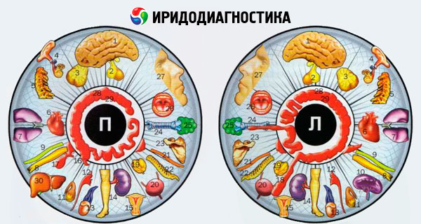
The map is a so-called hint for the doctor. After all, thanks to it, it is easy to determine which organs a person has problems with, as well as prevent the development of many diseases. In this regard, iridology is famous for its quick and accurate diagnosis.
Cost of iridology
What can be said about the cost of iridology and is the procedure generally available? In fact, anyone can use this method to determine the presence of diseases.
It is impossible to say that the procedure is affordable. But it is also impossible to say that it is expensive. Thus, on average, the cost of such an examination of the body fluctuates between 1800-2500 rubles, but this applies to Russia and its regions.
If we talk about Ukraine, such diagnostics will cost 350-400 hryvnia. This is the average price. Much depends on the region, the experience of the specialist and the clinic itself. Naturally, in the capital and cities close to it, the price will be much higher. Therefore, it is worth considering this diagnostics based on personal capabilities.
Again, for such a procedure, the price is not high at all. Because if you undergo all the standard examinations and tests, the cost will increase several times. This fact is still worth considering. Today, iridology is a new direction that will soon earn universal recognition.
[ 6 ]