Medical expert of the article
New publications
Stevens-Johnson syndrome
Last reviewed: 04.07.2025

All iLive content is medically reviewed or fact checked to ensure as much factual accuracy as possible.
We have strict sourcing guidelines and only link to reputable media sites, academic research institutions and, whenever possible, medically peer reviewed studies. Note that the numbers in parentheses ([1], [2], etc.) are clickable links to these studies.
If you feel that any of our content is inaccurate, out-of-date, or otherwise questionable, please select it and press Ctrl + Enter.

Causes Stevens-Johnson syndrome
Possible causes of the disease are often drugs (antibiotics, sulfonamides, analgesics, barbiturates, etc.). The disease can also occur due to other allergens. Currently, most dermatologists believe that) the basis of multiform exudative erythema and Stevens-Johnson syndrome and Lyell's syndrome are toxic-allergic reactions. Clinically and pathogenetically, the difference between them is not qualitative, but quantitative. In this case, the antigen-antibody reaction is directed at keratinocytes with the formation of circulating immune complexes in the blood serum, deposition of IgM and C3 complement component along the basement membrane of the epidermis and the upper part of the dermis.
Symptoms Stevens-Johnson syndrome
The disease is characterized by a rapid onset with pronounced general symptoms (high fever, arthralgia, myalgia). Then, after a few hours or 2-3 days, lesions of the skin and mucous membranes appear.
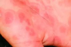
On the skin of the trunk, upper and lower extremities, face, genitals there appear disseminated rounded erythemato-edematous spots of a crimson color with a peripheral zone and a sunken cyanotic center with a diameter of 1 to 3-51 cm. Such elements of the rash resemble rashes of multiforme exudative erythema. Then, in the center of many of them, thin-walled flaccid blisters with serous or hemorrhagic contents are formed. The blisters, merging, reach enormous sizes. Opening, they leave juicy bright red painful erosions, along the edges of which there are fragments of the blister covers ("epidermal collar"). Under the influence of a light touch, the epidermis "slides" (positive Nikolsky symptom). The surface of the erosion over time becomes covered with yellowish-brown or hemorrhagic crusts.
On the mucous membrane of the oral cavity and eyes there is hyperemia, edema, flaccid blisters appear, after opening of which painful large erosions are formed. The red border of the lips is sharply edematous, hyperemic, has bleeding cracks and is covered with crusts. Often there are phenomena of blepharoconjunctivitis up to panophthalmia, damage to the mucous membrane of the urethra, bladder, upper respiratory tract. Severe general phenomena (fever, malaise, headaches, etc.) continue for 2-3 weeks.
Diagnostics Stevens-Johnson syndrome
In the blood, leukocytosis, eosinopenia, increased ESR, increased bilirubin, urea, aminotransferase levels, changes in plasma fibrinolytic activity due to activation of the proteolytic system, and a decrease in the total amount of protein due to albumins are observed.
What do need to examine?
How to examine?
What tests are needed?
Differential diagnosis
Differential diagnosis is carried out with pemphigus, Lyell's syndrome, etc.
 [ 20 ]
Who to contact?
Treatment Stevens-Johnson syndrome
Corticosteroids are prescribed at a rate of 1 mg/kg of the patient's weight (in terms of prednisolone) until a pronounced clinical effect is achieved, followed by a gradual reduction in the dose until complete withdrawal within 3-4 weeks. If oral administration is impossible, corticosteroids are prescribed parenterally. Measures are also taken to inactivate and remove antigens and circulating immune complexes from the body, using enterosorbents, hemosorption, plasmapheresis. To combat endogenous intoxication syndrome, 2-3 liters of fluid per day are administered parenterally (saline, hemodesis, Ringer's solution, etc.), as well as albumin and plasma. Calcium, potassium, antihistamines are also prescribed, and in the event of a threat of infection - broad-spectrum antibiotics. Externally, corticosteroid creams (lorinden C, dermovate, advantan, triderm, celestoderm with garomycin, etc.), 2% aqueous solutions of methylene blue, gentia violet are used.

