Medical expert of the article

New publications
Lung spirometry: what this procedure is, how it is performed
Last reviewed: 03.07.2025

All iLive content is medically reviewed or fact checked to ensure as much factual accuracy as possible.
We have strict sourcing guidelines and only link to reputable media sites, academic research institutions and, whenever possible, medically peer reviewed studies. Note that the numbers in parentheses ([1], [2], etc.) are clickable links to these studies.
If you feel that any of our content is inaccurate, out-of-date, or otherwise questionable, please select it and press Ctrl + Enter.
Evaluation of the external respiratory function is an integral component of a comprehensive clinical examination of a patient with pulmonary diseases. During anamnesis and physical examination, signs of respiratory function disorders of the lungs are identified, and then the degree of expression of these changes is purposefully assessed using standardized methods.
Spirometry is a method of measuring lung volume during various breathing maneuvers (calm breathing, maximum inhalation and exhalation, forced exhalation, maximum ventilation). Currently, volumes are measured based on air flow measurements - pneumotachometry (pneumotachography) with subsequent automatic data processing. The most common are recording calm deep inhalation and exhalation and evaluating forced exhalation parameters.
Other names for the method: forced expiratory flow-volume curve recording, Votchal-Tiffeneau test, forced expiratory spirography, pneumotachography with integration.
Currently, the use of such devices is unacceptable. Pneumotachometers determine air flow by measuring the pressure difference using differential manometers (Fleisch, Lily or Pitot tubes) or using "turbines" - inertialess propellers with light blades, while the patient breathes the surrounding air. The patient's lips and oral cavity are in contact only with a disposable mouthpiece.
Goals
- Diagnosis of disorders of the ventilation function of the lungs.
- Identification of the type (obstruction, restriction) and severity of the disorder.
- Evaluation of the course of pulmonary disease and the effectiveness of therapy (etiotropic, pathogenetic, in particular, bronchodilator).
- Evaluation of the reversibility of obstruction after inhalation of short-acting bronchodilators and evaluation of the response to provocative tests (methacholine, allergens).
- Determining the possibility of surgical treatment and assessing the postoperative condition.
- Objectification of the condition (for medical and social examination).
- Predicting the course of the disease.
Indications for the procedure
- Presence of complaints from the respiratory system.
- Changes in the respiratory organs on an X-ray (or other diagnostic methods).
- Disturbances in gas exchange (hypoxemia, hypercapnia, decreased saturation) and changes in laboratory parameters (polycythemia).
- Preparation for invasive examination or treatment methods ( bronchoscopy, surgery).
- Referral for medical and social examination.
 [
[ Preparation
The study is performed on an empty stomach or after a light breakfast. The patient should not take medications that affect the respiratory system (short-acting inhaled bronchodilators, cromoglycic acid for 8 hours, aminophylline, short-acting oral β 2 -adrenergic agonists for 12 hours, tiotropium bromide, long-acting inhaled and oral β 2 -adrenergic agonists, leukotriene receptor blockers for 24 hours, nedocromil and prolonged forms of theophylline for 48 hours, second-generation antihistamines for 72 hours), drink tea, coffee, or caffeinated beverages. Before the study, ties, belts, and corsets should be loosened, lipstick should be removed, and dentures are not recommended. Smoking is prohibited one hour before the procedure. If the examination is carried out in cold weather, the patient should warm up for 20-30 minutes.
Technique spirometry
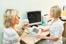
The spirometer is calibrated daily with the supplied 1-3 liter syringe (the "gold" standard is a three-liter syringe with a volume error of no more than 0.5%). Before the examination, the patient is explained the stages of the procedure, and maneuvers are demonstrated using a mouthpiece. During the procedure, the operator comments on the maneuver and directs the patient's actions.
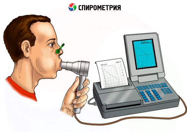
First, the vital capacity of the lungs is determined on inspiration (VC in ) or expiration (VC exp ). The nasal passages are blocked with a nasal clamp, the patient places the mouthpiece of the device (mouthpiece) into the oral cavity and tightly grasps it from the outside with his teeth. This ensures that the mouth is open during maneuvers. The patient's lips should tightly grasp the tube from the outside, preventing air leakage (this may be difficult for the elderly and for people with facial nerve damage). The patient is asked to breathe calmly through the mouth for adaptation (at this time, the spirometer calculates the tidal volume, respiratory rate, and minute respiratory volume, which are practically not used today). Then the patient is asked to calmly inhale deeply and calmly exhale deeply at least three times in a row. The patient should not make sharp inhalations or exhalations. The maximum amplitude of breathing from full exhalation to full inhalation is VC in, and from full inhalation to full exhalation is VC exp. During this procedure, a spirogram (a recording of changes in volume over time) is observed on the screen or display.
To record forced exhalation, the spirometer is switched to the appropriate mode and a flow-volume curve test is performed (recording the volumetric speed relative to the exhalation volume). The patient takes a calm, deep breath, holds his breath during inhalation and then exhales sharply with maximum effort and complete expulsion of air from the chest. The beginning of the exhalation should have a push character.
Only a correctly recorded curve with a distinct peak in the area no later than 25% from the start of the forced vital capacity (FVC) recording has practical significance: the peak of the expiratory flow rate should be no later than 0.2 s from the start of the forced exhalation. The duration of the forced exhalation should be at least 6 s, the end of the curve should have the form of a "plateau", during the recording of which the air flow is minimal, but the subject continues to exhale with effort.
At least three attempts are made to record forced expiration. The two attempts with the best results should not differ in FVC and forced expiratory volume in the first second (FEV 1 ) values by more than 150 ml.
Contraindications to the procedure
- Hemoptysis or pulmonary hemorrhage.
- Insufficiency of venous valves of the lower extremities with varicose veins, trophic disorders and a tendency towards increased blood clotting.
- Uncontrolled hypertension (systolic blood pressure >200 mmHg or diastolic blood pressure >100 mmHg).
- Aortic aneurysm.
- A history of myocardial infarction (or stroke) within the last 3 months.
- Postoperative period (one month after chest and abdominal surgery).
- Pneumothorax.
Normal performance
VC (FVC). FEV1 , peak expiratory flow rate (PEF) and instantaneous forced expiratory flow rates at 25%, 50% and 75% from the beginning of the FVC curve (MEF25, MEF50, MEF75) are expressed in absolute values (liters and liters per second) and as a percentage of the predicted values. The device calculates the norms automatically using regression equations based on the patient's gender, age and height. For VC (FVC). FEV1 , PEF the minimum normal value is 80% of the predicted value, and for MEF25, MEF50, MEF75 it is 60% of the predicted value. MEF25-75 is the average forced expiratory flow rate in the middle half of the FVC (i.e. between 25% and 75% of the FVC). COC25-75 reflects the condition of the small airways and is more significant than FEV1 in detecting early airway obstruction. COC25-75 is an effort-independent measure.
An isolated decrease in VC indicates the prevalence of restrictive disorders, and a decrease in FEV1 and the FEV1/FVC ratio ( or FEV1 / VC) indicates the presence of bronchial patency disorders or obstruction.
Based on the ratio of the main indicators, a conclusion is formulated.