Medical expert of the article

New publications
Small intestine (small bowel)
Last reviewed: 06.07.2025

All iLive content is medically reviewed or fact checked to ensure as much factual accuracy as possible.
We have strict sourcing guidelines and only link to reputable media sites, academic research institutions and, whenever possible, medically peer reviewed studies. Note that the numbers in parentheses ([1], [2], etc.) are clickable links to these studies.
If you feel that any of our content is inaccurate, out-of-date, or otherwise questionable, please select it and press Ctrl + Enter.
The small intestine (intestinum tenue) is a section of the digestive tract located between the stomach and the large intestine. The small intestine and the large intestine form the intestine, the longest part of the digestive system. The small intestine includes the duodenum, jejunum, and ileum. In the small intestine, chyme (food gruel), processed by saliva and gastric juice, is exposed to intestinal and pancreatic juice, as well as bile. In the lumen of the small intestine, when the chyme is mixed, its final digestion and absorption of its breakdown products occur. Food residues move into the large intestine. The endocrine function of the small intestine is important. Endocrinocytes of its integumentary epithelium and glands produce biologically active substances (secretin, serotonin, motilin, etc.).
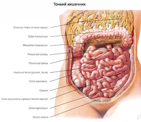
The small intestine begins at the level of the border of the bodies of the 12th thoracic and 1st lumbar vertebrae, ends in the right iliac fossa, is located in the region of the abdomen (middle region of the abdomen), reaching the entrance to the small pelvis. The length of the small intestine in an adult is 5-6 m. In men, the intestine is longer than in women, while in a living person, the small intestine is shorter than in a corpse, which has no muscle tone. The length of the duodenum is 25-30 cm; about 2/3 of the length of the small intestine (2-2.5 m) is occupied by the jejunum and about 2.5-3.5 m - the ileum. The diameter of the small intestine is 3-5 cm, it decreases towards the large intestine. The duodenum does not have a mesentery, unlike the jejunum and ileum, which are called the mesenteric part of the small intestine.
The jejunum and ileum make up the mesenteric portion of the small intestine. Most of them are located in the umbilical region, forming 14-16 loops. Some of the loops descend into the small pelvis. The loops of the jejunum are mainly located in the left upper part of the abdominal cavity, and the ileum in the right lower part of the abdominal cavity. There is no strict anatomical boundary between the jejunum and ileum. In front of the intestinal loops is the greater omentum, behind is the parietal peritoneum lining the right and left mesenteric sinuses. The jejunum and ileum are connected to the posterior wall of the abdominal cavity by means of the mesentery. The root of the mesentery ends in the right iliac fossa.
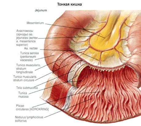
The walls of the small intestine are formed by the following layers: the mucous membrane with the submucosa, the muscular and outer membranes.
The mucous membrane (tunica mucosa) of the small intestine has circular (Kerckring's) folds (plicae circularis). Their total number reaches 600-700. The folds are formed with the participation of the submucosa of the intestine, their size decreases towards the large intestine. The average height of the folds is 8 mm. The presence of folds increases the surface area of the mucous membrane by more than 3 times. In addition to circular folds, longitudinal folds are characteristic of the duodenum. They are present in the upper and descending parts of the duodenum. The most pronounced longitudinal fold is located on the medial wall of the descending part. In its lower section, there is an elevation of the mucous membrane - a large papilla of the duodenum (papilla duodeni major), or the papilla of Vater. Here, the common bile duct and the pancreatic duct open through a common opening. Above this papilla, on the longitudinal fold, there is a small papilla of the duodenum (papilla duodeni minor), where the accessory pancreatic duct opens.
The mucous membrane of the small intestine has numerous outgrowths - intestinal villi (villi intestinales), there are about 4-5 million of them. On the area of 1 mm 2 of the mucous membrane of the duodenum and jejunum there are 22-40 villi, in the ileum - 18-31 villi. The average length of the villi is 0.7 mm. The size of the villi decreases towards the ileum. There are leaf-, tongue-, and finger-shaped villi. The first two types are always oriented across the axis of the intestinal tube. The longest villi (about 1 mm) have a predominantly leaf-shaped form. At the beginning of the jejunum, the villi usually have the shape of a tongue. Distally, the shape of the villi becomes finger-shaped, their length decreases to 0.5 mm. The distance between the villi is 1-3 µm. The villi are formed by loose connective tissue covered with epithelium. In the thickness of the villi there are many smooth myoocytes, reticular fibers, lymphocytes, plasma cells, eosinophils. In the center of the villi there is a lymphatic capillary (lactate sinus), around which there are blood vessels (capillaries).
The intestinal villi are covered on the surface with a single-layer high cylindrical epithelium located on the basal membrane. The bulk of the epithelial cells (about 90%) are columnar epithelial cells with a striated brush border. The border is formed by microvilli of the apical plasma membrane. On the surface of the microvilli is a glycocalyx represented by lipoproteins and glycosaminoglycans. The main function of columnar epithelial cells is absorption. The integumentary epithelium includes many goblet cells - single-celled glands that secrete mucus. On average, 0.5% of the cells of the integumentary epithelium are endocrine cells. In the thickness of the epithelium there are also lymphocytes penetrating from the stroma of the villi through the basal membrane.
In the spaces between the villi, intestinal glands (glandulae intestinales), or crypts, open onto the surface of the epithelium of the entire small intestine. In the duodenum, there are also mucous duodenal (Brunner's) glands of a complex tubular shape, located mainly in the submucosa, where they form lobules measuring 0.5-1 mm. Intestinal (Lieberkühn's) glands of the small intestine have a simple tubular shape, they occupy a place in the proper plate of the mucous membrane. The length of the tubular glands is 0.25-0.5 mm, the diameter is 0.07 mm. On an area of 1 mm 2 of the mucous membrane of the small intestine, there are 80-100 intestinal glands, their walls are formed by a single layer of epithelial cells. In total, there are more than 150 million glands (crypts) in the small intestine. Among the epithelial cells of the glands, columnar epitheliocytes with a striated border, goblet cells, intestinal endocrinocytes, borderless cylindrical (stem) cells and Paneth cells are distinguished. Stem cells are the source of regeneration of the intestinal epithelium. Endocrinocytes produce serotonin, cholecystokinin, secretin, etc. Paneth cells secrete erepsin.
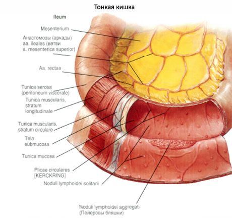
The lamina propria of the small intestinal mucosa is characterized by a large number of reticular fibers that form a dense network. The lamina propria always contains lymphocytes, plasma cells, eosinophils, and a large number of single lymphoid nodules (in children - 3-5 thousand).
In the mesenteric part of the small intestine, especially in the ileum, there are 40-80 lymphoid, or Peyer's, patches (noduli lymphoidi aggregati), which are clusters of single lymphoid nodules that are organs of the immune system. The plaques are located mainly along the antimesenteric edge of the intestine and have an oval shape.
The muscular plate of the mucous membrane (lamina muscularis mucosae) is up to 40 µm thick. It has an internal circular and external longitudinal layer. From the muscular plate, individual smooth myocytes extend into the thickness of the proper plate of the mucous membrane and into the submucosa.
The submucosa (tela submucosa) of the small intestine is formed by loose fibrous connective tissue. In its thickness are located branches of blood and lymphatic vessels and nerves, various cellular elements. The secretory sections of the duodenal (Brunper's) glands are located in the submucosa of the duodenum.
The muscular coat (tunica muscularis) of the small intestine consists of two layers. The inner layer (circular) is thicker than the outer (longitudinal) layer. The direction of the myocyte bundles is not strictly circular or longitudinal, but has a spiral course. In the outer layer, the spiral turns are more stretched compared to the inner layer. Between the muscular layers in the loose connective tissue are located the nerve plexus and vessels.
The serous membrane (tunica serosa) is located on the subserous base. It covers the small intestine on all sides, except for the duodenum, which is covered by the peritoneum only partially (in front), and in the remaining parts - by adventitia.
 [
[ Where does it hurt?
What do need to examine?
What tests are needed?