Medical expert of the article

New publications
Aortic wall and flail thickening
Last reviewed: 05.07.2025

All iLive content is medically reviewed or fact checked to ensure as much factual accuracy as possible.
We have strict sourcing guidelines and only link to reputable media sites, academic research institutions and, whenever possible, medically peer reviewed studies. Note that the numbers in parentheses ([1], [2], etc.) are clickable links to these studies.
If you feel that any of our content is inaccurate, out-of-date, or otherwise questionable, please select it and press Ctrl + Enter.
Among the pathologies of the vascular and circulatory systems, thickening of the aorta – the main arterial vessel – occupies one of the first places both in prevalence and in the severity of consequences.
What does aortic thickening mean? It is not a disease or a symptom of a disease, but a pathological change that has occurred in the structure of the wall of this vessel and can be detected using medical imaging equipment.
As a result of such changes, the aortic wall becomes less elastic, and this can negatively affect the hemodynamic function of the aorta, which ensures the continuity of the flow of oxygenated blood through other arterial vessels.
Causes aortic seals
The key causes of aortic thickening (its walls) are associated with a disorder of lipoprotein metabolism – dyslipidemia and its consequence – the deposition of LDL (low-density lipoproteins) on the inner surface of blood vessels in the form of cholesterol plaques, that is, atherosclerosis.
The second most common cause of decreased elasticity of the aortic walls is considered by specialists to be arterial hypertension, primarily isolated systolic arterial hypertension. A gradual increase in the density of the endothelium, subendothelial and medial layers of the aortic walls with the formation of dense fibrous structures makes them more rigid. And this occurs, as is believed, due to the constant hydromechanical pressure of blood continuously moving through the vessel at an average speed of 50 cm / sec. and blood pressure of at least 120 mm Hg. Although it is precisely this cause-and-effect relationship between the development of hypertension and increased rigidity of the aortic walls that has recently been questioned and may have an inverse sequence.
Also, the vascular wall can partially lose elasticity as a result of:
- age-related fibrous involution of the tissues of the aortic walls;
- chronic inflammation of the aorta (aortitis), which develops with tuberculosis, syphilis and streptococcal infections;
- the presence of systemic autoimmune pathologies (rheumatoid arthritis, systemic scleroderma or lupus);
- genetically determined collagenopathy (connective tissue dysplasia) in the form of vascular syndrome with endothelial dysfunction.
Risk factors
Among the risk factors for thickening of the aortic walls, in addition to hereditary predisposition and congenital aortic heart defects, angiologists and cardiologists note:
- age factor;
- smoking, alcohol abuse, excessive physical activity;
- too much animal fat in the diet (which contributes to increased LDL levels);
- metabolic syndrome;
- diabetes mellitus.
An important risk factor for decreased vascular elasticity is copper deficiency in the body, which reduces the strength of cross-links in the molecules of fibrillar proteins elastin and collagen (which are the main components of vascular wall tissue).
 [ 11 ], [ 12 ], [ 13 ], [ 14 ], [ 15 ], [ 16 ], [ 17 ], [ 18 ], [ 19 ]
Pathogenesis
The pathogenesis of increased aortic density directly depends on its cause and lies in the structural features of the wall of this vessel.
The aorta is an elastic artery with three membranes: internal, middle and external. The internal membrane (intima) consists of large endothelial cells connected to each other. Next comes the subendothelial layer of amorphous collagen and elastin fibers and above it is the elastin membrane separating the intima from the middle membrane.
The middle layer of the aorta is an extracellular matrix containing collagen, myocytes (smooth muscle cells), glycosaminoglycans, fibroblast cells, the structural protein fibronectin, and various immune cells. The outer layer of the aorta is formed by elastin and collagen fibers.
It is this structure of the aorta walls that ensures its elasticity, strength and biomechanical properties that determine the hemodynamic functions of this blood vessel. During systole (contraction of the left ventricle of the heart), the aorta walls are able to take on the ejection of blood, while the vessel expands, and the stretching of the wall provides potential energy that allows maintaining blood pressure during the diastolic phase of the cardiac cycle, since during this time the aorta is passively compressed. And the elastic recoil of its walls helps to preserve the energy of myocardial contractions and smooth out the pulse wave created by the heart.
High blood pressure (arterial hypertension) causes constant tension on the walls of the aorta and, over time, loss of their elasticity.
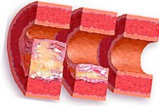
Sclerotic thickening of the aortic walls in atherosclerosis occurs due to the accumulation of lipids in the middle layer of its wall, which in the form of cholesterol conglomerates or cholesterol plaques are introduced directly into the intercellular matrix and gradually grow into the vessel, thickening its wall and reducing the lumen.
Also, the elastic layer of the aortic wall is subject to involutional changes, the pathogenesis of which is due to the fact that with age, its structural homogeneity is disrupted due to focal fibrosis or calcification deposits.
The increase in the level of fibronectin produced by the endothelial cells of the aortic membrane, which is typical of old age, not only leads to platelet aggregation and the formation of agglutination thrombi, but also activates the synthesis of growth factors (PDGF, bFGF, TGF) by the endothelium. As a result, the proliferation of fibroblasts and myocytes increases, and the aortic wall thickens and becomes denser.
As experts note, fibronectin levels can increase at any age with metabolic syndrome.
Symptoms aortic seals
A decrease in the elasticity of the aortic wall at an early stage of the pathological process does not manifest itself in any way. Moreover, aortic compaction on fluorography is often detected spontaneously - in the complete absence of any complaints from patients.
In addition, the symptoms of aortic compaction are non-specific. For example, moderate compaction of the aorta in the area of its arch may be accompanied by frequent headaches, dizziness, and increased fatigue.
When the aortic root and its ascending part become compacted, there is a feeling of discomfort in the mediastinum, increased heart rate, and pain behind the breastbone during physical exertion. Attacks similar to angina may occur if compaction of the aortic valve is combined with left ventricular hypertrophy.
With compaction of the abdominal aorta, patients may complain of weight loss, digestive problems, abdominal pain of a pulling nature, cramps in the muscles of the lower extremities, pain in the legs when walking and unilateral lameness.
Forms
The aorta is the main artery of the systemic circulation. It originates from the left ventricle of the heart, extending to the abdominal cavity, where it divides into two smaller (iliac) arteries. Specialists determine the forms or types of aortic compaction by its location.
If an increase in the density of the vascular wall is detected at the beginning of the aorta - in the area of its expanded (bulbar) part, then this is defined as compaction of the aortic root.
In the same part, next to the mouth of the vessel, there is the ascending aorta (no more than 5-6 cm long), originating in the chest on the left - near the lower edge of the third intercostal space, rising to the second rib on the right of the chest. With this localization, compaction of the ascending aorta is noted.
Additionally, because the ascending aorta extends from the heart's aortic valve, which regulates the flow of blood into the aorta from the left ventricle (and prevents backflow of blood), thickening of the aortic valve may be present.
Aortic insufficiency is associated with thickening of the cusps (elastic locking structures) of the aortic valve. The anatomical and functional connection can manifest itself in such simultaneous vascular pathology as thickening of the walls and cusps of the aorta.
Also, compaction of the aorta and the cusps of the aortic and mitral valves may be detected. If the aortic valve of the heart separates the aorta from the left ventricle, then the mitral valve separates the left atrium from it and does not allow blood to flow in the opposite direction during systolic contraction (i.e., prevents regurgitation).
Thickening of the aortic arch means localization of the pathology in the area where the ascending part of this vessel at the level of the second rib makes a left and upward turn (above the left pulmonary artery and left bronchus). Three large arteries branch off from the arch itself: the brachiocephalic trunk, the left common carotid and the left subclavian arteries.
The abdominal aorta is part of the descending aorta; it is located under the diaphragm. And compaction of the abdominal aorta can disrupt normal blood flow through the arteries that branch off from it – the iliac and mesenteric.
When compaction of the aorta and left ventricle (in the sense of its walls) is established, it means that prolonged high blood pressure in the patient has led to hypertrophy of the left ventricle (increase in the thickness of its wall) with simultaneous damage to the aortic wall of any etiology. Considering all the negative consequences of such a combination for hemodynamics, cardiologists note its danger: the mortality rate is 35-38 cases per thousand.
Complications and consequences
Is aortic thickening dangerous and what are the risks? Aortic thickening is a pathological condition of the vascular system that has certain consequences and complications, including life-threatening ones.
On the one hand, damage to the aorta by cholesterol plaques narrows the lumen of the vessel and reduces the elasticity of its walls, and on the other hand, causes compaction and expansion of the aorta - aneurysm. At the same time, constantly high blood pressure on the walls of the aorta can lead to their dissection, which is fraught with perforation of the vascular wall with huge blood loss and a fatal outcome.
Read also – Aneurysm of the abdominal aorta
Thickening of the aorta and aortic valve cusps leads to its insufficiency with diastolic regurgitation of part of the blood into the ventricle, which increases its volume and increases pressure during diastole. As a result, left ventricular hypertrophy develops, which can progress and cause a violation of its contractile functions.
The consequence of severe cases with compaction of a significant part of the aorta is a violation of coronary blood flow and myocardial ischemia, sometimes irreversible.
Diagnostics aortic seals
In order to identify pathology of the aortic walls - if the patient does not have a history of atherosclerosis or metabolic syndrome - blood tests for sugar and cholesterol should be taken.
Doctors can detect aortic thickening on fluorography (chest X-ray); aortic thickening is clearly visualized on cardiac ultrasound.
In addition, instrumental diagnostics uses:
- electrocardiography (ECG);
- ultrasound echocardiography;
- angiography with contrast agent;
- MRI.
Who to contact?
Treatment aortic seals
When the aortic walls are thickened, treatment is determined by the causes of this pathology. Thus, in atherosclerosis with damage to the aortic walls by cholesterol plaques, drugs are used that help reduce the level of cholesterol in the blood and reduce its production in the body, for more details see - Treatment of high cholesterol, and also - How to reduce cholesterol in the blood without drugs?
For any etiology of decreased elasticity of the aortic walls, vitamins C, E, B5 and PP are recommended, as well as polyunsaturated fatty acids omega-3 and omega-6.
In cases where the specific cause of the pathology is not established, the patient – provided there are no symptoms – is given standard advice: maintain a healthy lifestyle, eat right and avoid stress.
Surgical treatment is carried out:
- in case of aortic dissection – by stenting the vessel at the damaged area or by endoprosthetics;
- in case of compaction of the aortic and mitral valve cusps – their plastic correction or complete replacement;
- in case of aneurysm – resection with replacement of the removed area with a prosthesis.
Folk remedies for aortic compaction
The most effective folk remedy is garlic oil. To prepare it, you need to peel and chop one large head of garlic and mix it with 200-250 ml of corn oil.
This mixture should be stirred periodically throughout the day, after which the container should be tightly closed and placed in a cool place for a week.
Garlic oil is taken one teaspoon three times a day (30-40 minutes before meals). One course of such treatment lasts three months, after which it is necessary to take a break for a month.
Forecast
The prognosis for aortic wall thickening, as well as its treatment, is determined by the causes of this pathology…
Fortunately, aortic rupture due to dissection and aneurysm does not occur very often, but even timely intervention does not save from death in 90% of cases.