Medical expert of the article
New publications
Sclerite
Last reviewed: 04.07.2025

All iLive content is medically reviewed or fact checked to ensure as much factual accuracy as possible.
We have strict sourcing guidelines and only link to reputable media sites, academic research institutions and, whenever possible, medically peer reviewed studies. Note that the numbers in parentheses ([1], [2], etc.) are clickable links to these studies.
If you feel that any of our content is inaccurate, out-of-date, or otherwise questionable, please select it and press Ctrl + Enter.

Scleritis is a severe, destructive, vision-threatening inflammation involving the deep layers of the episclera and the sclera. Scleral infiltrate is similar to episcleral. Often one, sometimes two or more areas of inflammation develop simultaneously. In severe cases, inflammation can cover the entire pericorneal area. Usually, inflammation develops against the background of general immune pathology in middle-aged women. In half of the cases, scleritis is bilateral.
Symptoms include moderate pain, hyperemia of the eyeball, lacrimation and photophobia. Diagnosis is clinical. Treatment is with systemic glucocorticoids, and immunosuppressants may be used.
Causes sclerite
Scleritis is most common in women aged 30-50 years, and many have connective tissue diseases such as rheumatoid arthritis, SLE, periarteritis nodosa, Wegener's granulomatosis, or relapsing polychondritis. Some cases are caused by infection. Scleritis most often involves the anterior segment and comes in 3 types: diffuse, nodular, and necrotizing (perforating scleromalacia).
The causes of scleritis are very diverse. Previously, the most common causes of scleritis were tuberculosis, sarcoidosis, syphilis. Currently, the leading role in the development of scleritis is played by streptococcal infection, pneumococcal pneumonia, inflammation of the paranasal sinuses, any inflammatory focus, metabolic diseases - gout, collagenoses. Some authors point to a connection between the occurrence of scleritis due to rheumatism and polyarthritis. Pathological processes in scleritis develop according to the type of bacterial allergy, sometimes have an autoimmune nature, which causes their persistent recurrent course. Trauma (chemical, mechanical) can also be the cause of sclera diseases. In endophthalmitis, panophthalmitis, there may be secondary damage to the sclera.
Thus, the causes of scleritis are as follows
- In almost 50% of cases, scleritis develops against the background of systemic diseases of the body. The most common diseases are rheumatoid arthritis, Wegener's granulomatosis, relapsing polychondritis and nodular polyarthritis.
- Post-surgical scleritis. The exact cause is unknown, but there is a clear relationship with underlying systemic diseases; it is most common in women. Scleritis typically appears within 6 months after surgery as an area of intense inflammation and necrosis adjacent to the surgical site.
- Infectious scleritis is most often caused by the spread of an infectious process from a corneal ulcer.
Scleritis may also be associated with traumatic injury, pterygium excision, beta radiation, or mitomycin C. The most common infectious agents are Pseudomonas aeruginosa, Strept. pneumoniae, Staph. aureus, and herpes zoster virus. Pseudomonas scleritis is difficult to treat, and the prognosis for this type of scleritis is poor. Fungal scleritis is rare.
Symptoms sclerite
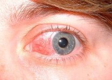
Scleritis begins gradually, over several days. Scleritis is accompanied by severe pain. The pain may radiate to other parts of the head. The eyeball is painful. The pain (often described as a deep, boring pain) is severe enough to interrupt sleep and affect appetite. Photophobia and lacrimation may occur. The affected areas are red with a purple tint, often surrounding the entire cornea ("ring scleritis"). Very often, scleritis is complicated by corneal diseases (sclerosing keratitis and inflammation of the iris and ciliary body). Involvement of the iris and ciliary body is expressed in the formation of adhesions between the pupillary margin of the iris and the lens, opacity of the aqueous humor of the anterior chamber, and deposition of precipitates on the posterior surface of the cornea. The conjunctiva is fused with the affected area of the sclera, the vessels cross in different directions. Sometimes scleral edema is detected.
The hyperemic patches occur deep beneath the bulbar conjunctiva and are purple in color compared to the hyperemia seen in episcleritis. The palpebral conjunctiva is normal. The involved area may be focal (i.e., one quadrant of the globe) or involve the entire globe and may contain a hyperemic, edematous, raised nodule (nodular scleritis) or an avascular area (necrotizing scleritis).
In severe cases of necrotizing scleritis, perforation of the globe may occur. Connective tissue disease occurs in 20% of patients with diffuse or nodular scleritis and in 50% of patients with necrotizing scleritis. Necrotizing scleritis in patients with connective tissue disease signals an underlying systemic vasculitis.
Necrotizing scleritis - most often occurs with inflammation, less often - without an inflammatory reaction (perforating scleromalacia).
Necrotizing scleritis without an inflammatory reaction often occurs against the background of long-standing rheumatoid arthritis, and is painless. The sclera gradually becomes thinner and protrudes outward. The slightest injury easily causes a rupture of the sclera.
Posterior scleritis is rare. Patients complain of pain in the eye. They have eye strain, sometimes limited mobility, exudative retinal detachment and optic disc edema may develop. Ultrasound and tomography can reveal thinning of the sclera in the posterior part of the eye. Posterior scleritis usually begins with general diseases of the body (rheumatism, tuberculosis, syphilis, herpes zoster) and is complicated by keratitis, cataracts, iridocyclitis, and increased intraocular pressure.
Deep scleritis is chronic and recurrent. In mild cases, the infiltrate resolves without severe complications.
With massive infiltration in the affected areas, necrosis of the scleral tissue and its replacement with scar tissue with subsequent thinning of the sclera occur. In places where there were areas of inflammation, traces always remain in the form of grayish zones as a result of thinning of the sclera, through which the pigment of the choroid and ciliary body shines through. As a result, stretching and protrusion of these areas of the sclera (staphyloma of the sclera) is sometimes observed. Vision deteriorates due to astigmatism developing as a result of the protrusion of the sclera and from the accompanying changes occurring in the cornea and iris.
 [ 9 ]
[ 9 ]
Where does it hurt?
What's bothering you?
Forms
Scleritis is classified according to anatomical criteria - anterior and posterior.
Among the anterior scleritis, the following clinical forms are distinguished: diffuse, nodular and the rarest - necrotizing.
 [ 10 ]
What do need to examine?
How to examine?
Who to contact?
Treatment sclerite
Primary therapy is systemic glucocorticoids (eg, prednisolone 1 mg/kg once daily). If the scleritis is tolerant to systemic glucocorticoids or the patient has necrotizing vasculitis and connective tissue disease, systemic immunosuppressive therapy with cyclophosphamide or azathioprine is indicated after consultation with a rheumatologist. If perforation is threatened, scleral tissue grafting may be indicated.
In the treatment, corticosteroids (drops dexanos, masidex, oftan-dexamethaeon or ointment hydrocortisone-POS), non-steroidal anti-inflammatory drugs in the form of drops (naklof), cyclosporine (cycloline) are used locally. Non-steroidal anti-inflammatory drugs (indomethacin, diclofenac) are also taken orally.
In necrotizing scleritis, which is considered an ocular manifestation of systemic diseases, immunosuppressive therapy (corticosteroids, cyclosporine, cytophosphamide) is necessary.

