Medical expert of the article

New publications
Red squamous lichen planus
Last reviewed: 04.07.2025

All iLive content is medically reviewed or fact checked to ensure as much factual accuracy as possible.
We have strict sourcing guidelines and only link to reputable media sites, academic research institutions and, whenever possible, medically peer reviewed studies. Note that the numbers in parentheses ([1], [2], etc.) are clickable links to these studies.
If you feel that any of our content is inaccurate, out-of-date, or otherwise questionable, please select it and press Ctrl + Enter.
Lichen planus is a common non-contagious inflammatory disease of the skin and mucous membranes, the course of which can be either acute or chronic.
The cause of this disease is still unknown.
 [ 1 ]
[ 1 ]
Epidemiology
The overall prevalence of lichen planus in the general population is approximately 0.1 - 4%. It occurs more frequently in women than in men, in a ratio of 3:2, and is diagnosed in most cases between the ages of 30 and 60 years.
[ 2 ]
Causes red flat shingles
The causes and pathogenesis of lichen planus have not been fully established. Lichen planus is a polyetiological disease that most often develops in connection with the use of medications, contact with chemical allergens, primarily with reagents for color photography, infections, especially viral ones, and neurogenic disorders. Lesions of the oral mucosa in lichen planus are often caused by hypersensitivity to components of dentures and fillings. There is evidence of a connection between the disease and liver diseases, carbohydrate metabolism disorders, autoimmune diseases, primarily lupus erythematosus.
There are theories of viral, infectious-allergic, toxic-allergic and neurogenic origin of the disease. In recent years, studies have shown that changes in the immune system are of great importance in the pathogenesis of lichen planus. This is evidenced by a decrease in the total number of T-lymphocytes and their functional activity, the deposition of IgG and IgM in the dermoepidermal border, etc.
Pathogenesis
In the typical form of lichen planus, the characteristic signs are hyperkeratosis with uneven granulosis, acanthosis, vacuolar dystrophy of the basal layer of the epidermis, diffuse strip-like infiltrate in the upper part of the dermis, closely adjacent to the epidermis, the lower border of which is "blurred" by the cells of the infiltrate. Exocytosis is noted. In the deeper parts of the dermis, dilated vessels and perivascular infiltrates are visible, consisting mainly of lymphocytes, among which are histiocytes, tissue basophils and melanophages. In old foci, the infiltrates are less dense and consist mainly of histiocytes.
The verrucous, or hypertrophic, form of lichen planus is characterized by hyperkeratosis with massive horny plugs, hypergranulosis, significant acanthosis, and papillomatosis. As with the typical form, in the upper part of the dermis there is a diffuse strip-like infiltrate of lymphoid cells, which, penetrating into the epidermis, seem to "blur" the lower border of the epidermis.
The follicular form of lichen planus is characterized by a sharp widening of the mouths of the hair follicles, filled with massive horny plugs. Hair is usually absent. The granular layer is thickened, there is a dense lymphocytic infiltrate at the lower pole of the follicle. Its cells penetrate the epithelial sheath of the hair, as if erasing the boundary between it and the dermis.
The atrophic form of lichen planus is characterized by epidermal atrophy with smoothing of epithelial outgrowths. Hypergranulosis and hyperkeratosis are expressed less strongly than in the usual form. A strip-like infiltrate in the dermis is rare, more often it is perivascular or merging, consists mainly of lymphocytes, in the subdermal sections there is proliferation of histiocytes. It is always possible, although with difficulty, to find areas of "blurring" of the lower border of the basal layer by the infiltrate cells. Sometimes a significant number of melanophages with pigment in the cytoplasm are found among the infiltrate cells - a pigment form.
The pemphigoid form of lichen planus is characterized mostly by atrophic phenomena in the epidermis, smoothing of its outgrowths, although hyperkeratosis and granulosis are almost always expressed. In the dermis - a scanty, often perivascular infiltrate of lymphocytes with an admixture of a large number of histiocytes. In some areas, the epidermis peels off from the underlying dermis with the formation of cracks or fairly large blisters.
The coral-shaped form of lichen planus is characterized by an increase in the number of vessels, around which a focal lymphocytic infiltrate is detected. Hyperkeratosis and granulosis are expressed much less strongly, sometimes parakeratosis may be present. It is always possible to see in separate areas of epidermal outgrowths "blurring" of the lower border of the basal layer to vacuolization of its cells.
The histological picture of the lesion in lichen planus of the mucous membranes is similar to that described above, however, hypergranulosis and hyperkeratosis are absent; parakeratosis is more common.
Histogenesis of lichen planus
In the development of the disease, great importance is attached to cytotoxic immune reactions in the basal layer of the epidermis, since activated cytotoxic T-lymphocytes predominate in cellular infiltrates, especially long-existing elements. The number of Langerhans cells in the epidermis is significantly increased. RG Olsen et al. (1984) using an indirect immunofluorescence reaction found an antigen specific for lichen planus in both the spinous and granular layers of the epidermis. In an immunoelectron microscopic study of the pemphigoid form of C. Prost et al. (19?5) found deposits of IgG and the C3 component of complement in the lamina hicula of the basement membrane in the peribullous zone of the lesion, as in bullous pemphigoid, but unlike the latter, they are not in the roof of the bladder, but in the zone of the basement membrane along the bottom of the bladder. Familial cases of the disease indicate a possible role of genetic factors, which is also supported by the possibility of an association of lichen planus with some HLA histocompatibility antigens.
Histopathology of lichen planus
Histologically, hyperkeratosis, thickening of the granular layer with an increase in keratohyalin cells, uneven acanthosis, vacuolar degeneration of the cells of the basal layer, diffuse strip-like infiltrate of the papillary layer of the dermis, consisting of lymphocytes, much less often - histiocytes, plasma cells and polymorphonuclear leukocytes and closely adjacent to the epidermis with penetration of infiltrate cells into the epidermis (exocytosis) are characteristic.
Symptoms red flat shingles
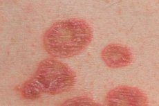
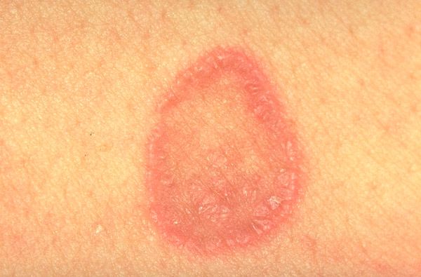
The disease often occurs in adults, mainly in women. The typical form of lichen planus is characterized by a monomorphic rash (from 1 to 3 mm in diameter) in the form of polygonal papules of a red-violet color with an umbilicated depression in the center of the element. On the surface of larger elements, Wickham's mesh is visible (opal-shaped white or grayish dots and stripes - a manifestation of uneven granulosis), which is clearly visible when the elements are lubricated with vegetable oil. Papules can merge into plaques, rings, garlands, and be located linearly. In the acute stage of dermatosis, a positive Koebner phenomenon is observed (the appearance of new rashes in the area of skin trauma). Rashes are usually localized on the flexor surfaces of the forearms, wrist joints, lower back, abdomen, but can also appear on other areas of the skin. The process can sometimes become widespread, up to universal erythroderma. Regression of the rash is usually accompanied by hyperpigmentation. The mucous membrane lesion may be isolated (oral cavity, genitals) or combined with skin pathology. Papular elements have a whitish color, a reticular or linear character and do not rise above the level of the surrounding mucous membrane. There are also warty, erosive-ulcerative forms of mucous membrane lesions.
The nail plates are affected in the form of longitudinal grooves, depressions, areas of clouding, longitudinal splitting and onycholysis. Subjectively, intense, sometimes excruciating itching is noted.
What's bothering you?
Forms
There are several clinical forms of the disease:
- bullous, characterized by the formation of blisters with serous-hemorrhagic contents on the surface of papules or against the background of typical manifestations of lichen planus on the skin and mucous membranes;
- annular, in which papules are grouped in the form of rings, often with a central zone of atrophy;
- warty, in which the rash is usually located on the lower extremities and is represented by dense warty plaques that are bluish-red or brown in color. Such lesions are very resistant to the therapy;
- erosive-ulcerative, occurring most often on the mucous membrane of the mouth (cheeks, gums) and genitals, with the formation of painful erosions and ulcers of irregular shape with a red velvety bottom. Typical papular elements are noted on other areas of the skin. It is observed more often in patients with diabetes mellitus and hypertension;
- atrophic, manifested by atrophic changes along with typical foci of lichen planus. Secondary atrophy of the skin is possible after the resolution of the elements, especially plaques;
- pigmented, manifested by pigment spots that precede the formation of papules, most often affecting the face and upper limbs;
- linear, characterized by linear lesions;
- psoriatic, manifested in the form of papules and plaques covered with scales that have a silvery-white color, as in psoriasis.
The usual form of lichen planus is characterized by rashes of small shiny papules of polygonal outlines, red-violet color with a central umbilicated pit, located mainly on the flexor surface of the limbs, trunk, on the mucous membrane of the oral cavity, genitals, often grouped in the form of rings, garlands, semi-arcs, linear and zosteriform foci. In the mucous membrane of the oral cavity, along with typical rashes, exudative-hyperemic, erosive-ulcerative and bullous ones are noted. Peeling on the surface of the papules is usually insignificant, the scales are separated with difficulty, psoriasiform peeling is occasionally observed. After lubricating the nodules with vegetable oil, a mesh-like pattern (Wickham's mesh) can be found on their surface. Changes in the nails in the form of longitudinal striations and cracks in the nail plates are often encountered. In the active phase of the process, a positive Koebner symptom is observed and, as a rule, there is itching of varying intensity.
The course of the disease is chronic, only in rare cases is an acute onset observed, sometimes in the form of a polymorphic rash merging into large foci up to erythroderma. With a long-term existence of the process, especially with localization on the mucous membranes, warty and erosive-ulcerative forms, the development of cancer is possible. Combinations of lichen planus and discoid lupus erythematosus with localization of foci mainly on the distal parts of the extremities, having histological and immunomorphological signs of both diseases, have been described.
The warty, or hypertrophic, form of lichen planus is much less common and is clinically characterized by the presence of sharply defined plaques with a warty surface, pronounced hyperkeratosis, significantly elevated above the skin surface, accompanied by intense itching on the anterolateral surfaces of the shins, and less often on the hands and other areas of the skin. Around these lesions, as well as on the mucous membrane of the oral cavity, rashes typical of lichen planus may be detected.
The vegetative form differs from the previous one by the presence of papillomatous growths on the surface of the lesions.
The follicular, or pointed, form is characterized by rashes of pointed follicular nodules with a horny plug on the surface, in place of which atrophy and baldness may develop, especially when the rash is localized on the head (Graham-Little-Piccardi-Lassuer syndrome).
The atrophic form is characterized by the presence of atrophy at the site of regressing, predominantly ring-shaped rashes. Along the edge of the atrophic elements, a brownish-blue rim of preserved ring infiltration can be seen.
The pemphigoid form of lichen planus is the rarest and is clinically characterized by the development of vesicular-bullous elements with transparent contents, usually accompanied by itching. Bullous lesions are located in the area of papular rashes and plaque lesions, as well as against the background of erythema or clinically healthy skin. This form may be paraneoplasia.
The coral-shaped form is observed very rarely, clinically characterized by rashes mainly on the neck, in the shoulder girdle, on the chest, abdomen of large flattened papules, located in the form of beads, reticular, in the form of stripes. Around such foci, typical rashes can be observed, often hyperpigmented. A.N. Mehregan et al. (1984) do not consider this form a variety of lichen planus. They believe that this is probably an abnormal reaction of the skin to trauma, manifested by the formation of linear hypertrophic scars.
"Keratosis lichenoides chronica" described by M.N. Margolis et al. (1972) and manifested by rashes on the scalp and face similar to seborrheic dermatitis, as well as lichenoid hyperkeratotic papules on the skin of the extremities, is considered as a variant of lichen planus, similar in clinical signs to the coral form. Three types of hyperkeratotic lesions are indicated, which are observed in most patients:
- linear, lichenoid and warty;
- yellow keratotic lesions and
- slightly raised papules with horny plugs.
Frequent involvement of the palms and soles in the form of diffuse keratosis and individual hyperkeratotic papules is noted; sometimes the nails are affected, they thicken, turn yellowish, and longitudinal ridges appear on their surface. According to A.N. Mehregan et al. (1984), this clinical form corresponds not to coral-shaped, but to warty red flat lichen.
Ulcerative form of lichen planus is also very rare. Ulcerative lesions are painful, especially when localized on the lower extremities, they are small in size with infiltrated edges, reddish-bluish in color. At the same time, typical rashes for lichen planus can be found on other areas of the skin.
The pigmented form of lichen planus may manifest itself in the form of nodular elements typical in morphology, but having a brownish-brown color, diffuse foci of pigmentation, changes similar to poikilodermic, in which nodular rashes can be hardly detected. In some cases, typical manifestations of lichen planus are found on the mucous membrane of the oral cavity. Some authors consider persistent dyschromic erythema, or "ash dermatosis", a variant of the pigmented form of lichen planus, clinically manifested by multiple ash-gray spots located mainly on the neck, shoulders, back, not accompanied by subjective sensations.
The subtropical form is found mainly in the Middle East, characterized by pigmented ring-shaped lesions, located mainly on exposed parts of the body. Itching is insignificant or absent, nails and scalp are rarely affected.
The course of lichen planus is usually chronic. Elements on the mucous membranes regress more slowly than on the skin. Long-standing hypertrophic and erosive-ulcerative lesions can transform into squamous cell carcinoma.
[ 7 ]
What do need to examine?
How to examine?
What tests are needed?
Differential diagnosis
Differential diagnosis of lichen planus is carried out with:
- psoriasis,
- nodular pruritus,
- lichenoid and warty tuberculosis of the skin.
Who to contact?
Treatment red flat shingles
The therapy depends on the prevalence, severity and clinical forms of the disease, as well as on concomitant pathology. Drugs affecting the nervous system (bromine, valerian, motherwort, elenium, seduxen, etc.), hingamine drugs (delagyl, plaquepil, etc.), as well as antibiotics (tetracycline series), vitamins (A, C, B, PP, B1, B6, B22,) are used. In widespread forms and in severe cases, aromatic retinoids (neotigazon, etc.), corticosteroid hormones, PUVA therapy (Re-PUVA therapy) are prescribed.
Externally, antipruritic agents (shaken suspensions with anesthesin, menthol), ointments with corticosteroid hormones (elokom, betnovate, dermovate, etc.), often applied under an occlusive dressing, are prescribed; warty lesions are injected with hingamine or diprospan; when treating mucous membranes, 1% dibunol ointment is used, rinses with infusions of sage, chamomile, eucalyptus.