Medical expert of the article

New publications
Psoriasis spots: red, white, pigmented spots
Last reviewed: 04.07.2025

All iLive content is medically reviewed or fact checked to ensure as much factual accuracy as possible.
We have strict sourcing guidelines and only link to reputable media sites, academic research institutions and, whenever possible, medically peer reviewed studies. Note that the numbers in parentheses ([1], [2], etc.) are clickable links to these studies.
If you feel that any of our content is inaccurate, out-of-date, or otherwise questionable, please select it and press Ctrl + Enter.
Causes psoriasis spots
Although the key causes of the formation of spots in psoriasis have not been finally established, modern dermatology adheres to the most convincing version - the autoimmune nature of hyperproliferation and abnormal differentiation of keratin cells of the skin. This is confirmed not only by the presence of psoriasis in family histories, but also by the identified links between disorders in this pathology and aberrations of the PSORS genetic loci on chromosomes 12.
Thus, the strongest correlation was established for the PSORS-1 locus on the short arm of chromosome 6 in the 6p21.3 region, where genes encoding proteins that control the response of immune system cells to foreign genes and provide the functions of the human leukocyte antigen (HLA) are concentrated.
The most important function of human skin is the immune function, therefore, as a result of genetic factors, a hypertrophied reaction of local immunity develops, which manifests itself in the formation of spots in psoriasis. The local protective reaction begins with the synthesis of a complex of cytokines - inflammation mediators - by T- and B-lymphocytes, macrophages, mast cells, neutrophils, histiocytes, basophils: prostaglandins (E1, E2, T2a); interleukins IL-5, IL-6, IL-8; leukotrienes; tumor necrosis factor alpha (TNFα), which stimulates the formation of an inflammatory focus; transforming growth factor alpha (TGFα), etc.
In addition, the keratinocytes themselves, being included in the intracellular autoimmune process activated by cytokines, begin to synthesize interleukins (IL-1α and IL-1β) that initiate increased cell growth; epidermal growth factor (EGF), which increases the rate of protein synthesis; and nerve growth factor (NGF), which promotes cell proliferation.
As a result, all this repeatedly increases the expression of basal keratinocytes and the rate of their migration to the upper layers of the epidermis, which disrupts the physiological process of keratinization (keratinization) in certain areas of the skin. This is exactly how the pathogenesis of the appearance of spots in psoriasis appears today - localized thickening of the stratum corneum of the skin. Its lamellar exfoliation (desquamation) on the surface of the rash is caused by accelerated keratinization of keratinocytes. And as a result of the activation of the endings of sensitive nerve C-fibers of the skin, the production of neuropeptides, substance P and vasodilator calcitonin polypeptide CGRP increases, which cause persistent hyperemia of the rash - red spots in psoriasis.
 [ 4 ]
[ 4 ]
Symptoms
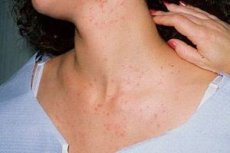
Psoriasis spots have several modifications and corresponding symptoms. In the most common form of psoriasis vulgaris, spots look like plaques that first appear as flat, clearly demarcated erythematous macules – round-oval, less than 1 cm in diameter – or dense red papules that rise slightly above healthy skin. They usually appear on the elbows, knees, lower back and head (on the scalp), and then on any other parts of the body, but almost always symmetrically.
Increasing in size at the progressive stage of the disease, these red spots in psoriasis can merge to form plaques up to several centimeters in diameter. Some red spots are bordered by a "halo" of pale skin (Voronov's ring). Dermatologists see the etiology of this symptom in the release of substances into the blood that inhibit the increase in the level of prostaglandins, which dilate the capillaries of the skin. However, when the disease progresses, the rings surrounding the papules are pink in color and are the border of the inflammatory process zone.
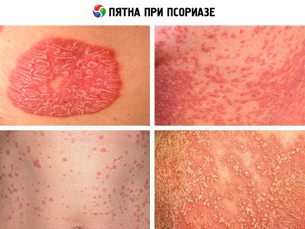
Quite quickly the spots become denser and more prominent, and their top is covered with silvery-white scales (keratinized skin cells, similar in appearance to stearin shavings). And such a plaque - a stearin spot in psoriasis - is a characteristic sign of the disease. By the way, as is the next symptom, which is an increase in desquamation after an attempt to scrape off the scales with a fingernail. Moreover, under the scraped off keratinized skin cells the patient sees a wet shiny border (terminal) film of an intensely pink color - the epidermis modified in structure. And here another symptom of spots in psoriasis appears - the Auspitz symptom in the form of protruding small drops of blood.
The appearance of the spots varies depending on the stage of psoriasis, and during the regression of the disease, the plaques decrease, become pale, flat and stop flaking. At this stage, in the place where the plaques had resolved, either discolored, almost white spots after psoriasis appear (due to the absence of the pigment melanin in the epidermis damaged by the autoimmune process), or darker pigment spots after psoriasis. In the latter case, the cause may be associated with a more active state of melanocytes (skin cells that produce pigment) in some patients, as well as with higher levels of pituitary melanocortin (MSH) and adrenocorticotropic hormone (ACTH).
Who to contact?
Diagnostics psoriasis spots
In addition to typical spots in psoriasis, rashes can have other morphological subtypes:
- very small hyperemic papules characteristic of punctate psoriasis;
- a rash in the form of small (2-10 mm) nodules with a drop-shaped form of the disease (most typical for children);
- ring-shaped spots with intact skin inside them (annular psoriasis, most often found in pediatric practice);
- red-orange spots covered not with scales, but with thickened multi-layered crusts of a dirty yellow color, under which weeping skin is exposed, occur with so-called exudative psoriasis;
- cone-shaped plaques 2-5 cm on the arms and legs (in the area of skin swelling near the joints) with severe hyperkeratosis, reminiscent of oyster shells, can be called rupioid psoriasis;
- If small pustules appear on a red psoriatic spot localized on the palms of the hands or soles of the feet, which dry up to purulent crusts; the skin is painful, and the inflamed area causes severe burning, then specialists diagnose pustular psoriasis. And if the affected area expands and there is fever, we may be talking about generalized pustular psoriasis.
[ 5 ]
Differential diagnosis
How psoriasis is diagnosed and why differential diagnostics is necessary – read more in the publication Vulgar psoriasis
Treatment psoriasis spots
Since systemic therapy for psoriasis is currently limited to long-term use of just a few drugs that have serious side effects (which will be discussed below), treatment of psoriasis spots with topical agents, that is, symptomatic treatment of psoriasis, is the most commonly used tactic for treating patients with this diagnosis.
We offer a detailed publication – Psoriasis, in which you will find a description of treatment methods, including physiotherapeutic treatment of psoriasis.
What needs and can be applied externally to reduce spots in psoriasis is described in detail in the articles - Creams for psoriasis and Non-hormonal ointments for psoriasis
And if local treatment does not improve the skin condition, then dermatologists have in their arsenal such drugs as Methotrexate, Cyclosporine and Acitretin.
Methotrexate is an immune-suppressing antimetabolite that may be prescribed (orally or by injection once a week) to adults with severe psoriasis or psoriatic arthritis. The drug helps reduce psoriasis symptoms within five to six weeks of starting treatment, but some people take Methotrexate for up to six months. Side effects include nausea, fatigue, headaches, and increased sensitivity to sunlight. There is also a significant risk of liver damage in patients taking Methotrexate, with about one in two hundred patients developing cirrhosis.
Cyclosporine is an immunosuppressant drug that inhibits the activity of immune cells by slowing the proliferation of keratinocytes. Typically, the drug provides some relief within a few weeks and achieves a stable level of rash control within three to four months. However, the use of Cyclosporine may increase the risk of kidney dysfunction, skin cancer, and other serious pathologies.
And the drug Acitretin (other trade name Neotigason) is a retinoid, a derivative of vitamin A, which is taken orally one capsule daily for two to four months. Possible side effects are expressed in the form of hypervitaminosis A (increased brittleness of nails, hair loss, peeling skin all over the body, muscle and joint pain, increased calcium levels in the blood, etc.).
Other retinoid drugs are recommended as an alternative – Isotretinoin (Accutane, Roaccutane) or Etretinate (Tigazon). The standard daily dose is 0.1 mg per kg of body weight; the maximum duration of treatment is 4 months (with a two-month break before the next course of treatment). Retinoids, like other systemic drugs used in the treatment of psoriasis, are absolutely contraindicated during pregnancy and lactation.
If you are interested in folk remedies for treating psoriasis spots, read the article – Treating Psoriasis at Home
And also learn about what can be Prevention of psoriasis