^

Health

Ultrasound diagnosis (ultrasound)

Ligament ultrasound

Ligaments are fibrillar structures that connect two bone structures together. There are two types of ligaments: intra-articular and extra-articular. This difference determines the differentiated approach to their study. Since ultrasound examination of intra-articular ligaments is difficult due to bone structures, MRI is used to evaluate them.

Tendon ultrasound

The ultrasound method competes with MRI in diagnosing tendon pathology. The main advantage of ultrasound is: high spatial resolution when scanning soft tissue structures and the possibility of dynamic research in real time.

Muscle ultrasound

About 30% of all sports injuries are caused by muscle tissue pathology. Ultrasound examination is the leading method in diagnosing muscle tissue pathology, surpassing magnetic resonance imaging in resolution.

Ultrasound of skin and subcutaneous fatty tissue

Skin thickness varies depending on the location, being thicker in men than in women. The subcutaneous fat layer usually appears hypoechoic with alternating hyperechoic thin fibers reflecting connective tissue layers.

Ultrasound-guided biopsy

Not every effusion or abscess needs to be punctured under ultrasound guidance, but it is important to use echography when puncture formations located near vital organs. Ultrasound is used to select the shortest distance to the object and the safest trajectory for the needle to pass.

Pleural ultrasound

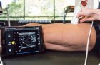
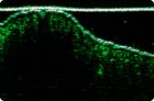
Pleural effusion is hypoechoic or moderately echogenic, sometimes with thick septa. Liquid blood and pus are also anechoic, but septa may produce reflections. It is not always possible to differentiate fluid from solid pleural or peripheral lung lesions. Turn the patient into different positions and repeat the examination.

Pericardial ultrasound

Fluid around the heart is visualized as an anechoic band around the heart muscle. (Anterior anechoic fat can simulate fluid.) If a small amount of fluid is present, the shape of the band may vary depending on the phase of the cardiac cycle.

Neck ultrasound

Indications for ultrasound examination of the neck: Palpable formation in the neck area. Pathology of the carotid arteries (rough noise, symptoms of insufficiency). In this case, a Doppler study is necessary to establish an accurate diagnosis.

Hip ultrasound in newborns

Certain skills and abilities are required to perform ultrasound examinations of the hip joints of newborns in order to exclude congenital hip dislocations.

Neurosonography

Neurosonography is a section of ultrasound diagnostics that deals with the study of the brain in newborn children. It is currently an integral part of traditional examination in neonatology and perinatal neurology, without which no examination by a pediatric neurologist and/or neonatologist is complete.

Pages

You are reporting a typo in the following text:
Simply click the "Send typo report" button to complete the report. You can also include a comment.