Medical expert of the article

New publications
Pineocytoma of the pineal gland.
Last reviewed: 12.07.2025

All iLive content is medically reviewed or fact checked to ensure as much factual accuracy as possible.
We have strict sourcing guidelines and only link to reputable media sites, academic research institutions and, whenever possible, medically peer reviewed studies. Note that the numbers in parentheses ([1], [2], etc.) are clickable links to these studies.
If you feel that any of our content is inaccurate, out-of-date, or otherwise questionable, please select it and press Ctrl + Enter.
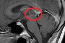
A well-localized brain tumor, pineocytoma of the pineal gland or pineal body (corpus pineale), occurs predominantly in adults. [ 1 ]
According to the WHO classification of CNS tumors based on histological features, pineocytoma is classified as a grade I tumor, i.e. it is a slowly growing benign formation with the possibility of cure after its removal. [ 2 ]
Causes pineocytomas
The underlying causes of most pineal gland tumors are unknown. From a morphological point of view, pineocytoma develops as a result of the proliferation of the main cells of the pineal gland parenchyma – pinealocytes. [ 5 ]
This tumor is considered well differentiated and belongs to the tumors of neuroectodermal tissue; it consists of small, cytologically benign mature cells similar to pinealocytes (arranged in the form of pineocytomatous pseudorosettes).
Since the pineal gland produces the hormone melatonin, which regulates the body's day and night cycle (circadian rhythms), and functions as a neuroendocrine organ, the main function of pinealocytes is secretory. And experts have put forward a version of the neuroendocrine etiology of pineocytoma tumor cells. [ 6 ]
After all, pinealocytes are modified nerve cells: they have more mitochondria than normal neurons, and the activity of these cellular organelles changes cyclically throughout the day. [ 7 ]
Risk factors
Potential risk factors may include exposure to ionizing radiation or toxic substances, although many researchers are inclined to believe that this type of pineal gland tumor has a genetic etiology. And there is considerable evidence to support this. [ 8 ]
Pathogenesis
Most likely, the pathogenesis of pineal gland pineocytoma is associated with disturbances in intracellular processes, which may be caused by chromosomal rearrangements and changes in the expression of certain genes:
- located on the short arms of the X and Y chromosomes, the ASMT gene, which encodes acetylserotonin-O-methyltransferase, an enzyme of the pineal gland that catalyzes the final stage of melatonin biosynthesis;
- the SAG gene, expressed in the retina and pineal gland, which encodes the highly antigenic protein S-arrestin;
- located on the short arm of the X chromosome, the SYP gene encoding synaptophysin, an integral membrane glycoprotein of endocrine cells, which is activated in all tumors of neuroendocrine origin;
- S100B gene, which encodes cytoplasmic and nuclear S100 proteins involved in cell differentiation and cell cycle regulation. [ 9 ]
Symptoms pineocytomas
The onset of pineocytoma formation is asymptomatic, and its first signs in patients are manifested by attacks of headache and dizziness, as well as nausea and vomiting.
As the tumor grows, other symptoms appear:
- diplopia (double vision), problems with focusing, lack of pupillary response to light and impaired up-down eye movement - Parinaud's syndrome, which develops due to compression of the tectum (plate of the quadrigeminal plate) in the dorsal part of the midbrain (at the level of the superior colliculus and the third cranial nerve);
- involuntary rapid eye movements with retraction of the eyelids (convergence-retraction nystagmus);
- hand tremors and decreased muscle tone;
- vestibulo-ataxic syndrome – gait and coordination disorder.
Complications and consequences
The main complications and consequences are also caused by the gradual increase in the size of the neoplasm.
A pineocytoma can compress the aqueductus cerebri, disrupting the circulation of cerebrospinal fluid with an increase in its pressure and the development of hydrocephalic syndrome. Increased intracranial pressure can lead to seizures and be life-threatening.
If the tumor affects the thalamus, there may be one-sided weakness (hemiparesis) and loss of sensitivity; when a pineocytoma affects the hypothalamus, temperature control, water regulation, and sleep are disrupted.
Endocrine insufficiency of the pineal gland leads to insomnia, and when the tumor formed in childhood, there may be growth retardation, premature puberty, changes in body weight, and the development of diabetes insipidus. [ 10 ]
Diagnostics pineocytomas
Given the fact that the symptoms of the tumor are nonspecific, clinical diagnosis can only rely on tests, in particular, cerebrospinal fluid analysis, biopsy and imaging methods. [ 11 ]
Thus, instrumental diagnostics are carried out using magnetic resonance imaging (MRI) of the brain and the entire spine with contrast enhancement; ultrasound encephalography, cerebral angiography and ventriculography. [ 12 ]
Differential diagnosis
Differential diagnosis includes pineal cyst and malignant pineoblastoma, papillary tumor of the pineal gland, germinoma, embryonal carcinoma, choriocarcinoma, paraphyseal meningioma or cavernoma, teratoma, and pineal astrocytoma.
Who to contact?
Treatment pineocytomas
In case of pineocytoma of the pineal gland, surgical treatment is performed by removing it. [ 13 ]
Prevention
Since pineocytoma is a benign tumor, the prognosis after surgery is favorable: five-year survival after its total removal is 86-100%.