Medical expert of the article
New publications
Pineoblastoma of the brain
Last reviewed: 12.07.2025

All iLive content is medically reviewed or fact checked to ensure as much factual accuracy as possible.
We have strict sourcing guidelines and only link to reputable media sites, academic research institutions and, whenever possible, medically peer reviewed studies. Note that the numbers in parentheses ([1], [2], etc.) are clickable links to these studies.
If you feel that any of our content is inaccurate, out-of-date, or otherwise questionable, please select it and press Ctrl + Enter.

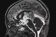
The formation of a blastoma, a rare type of cancerous neuroectodermal tumor, in the pineal or pineal endocrine gland of the brain is defined as pineoblastoma of the brain.
In the ICD-10 neoplasms section, this tumor has the code C75.3 (malignant neoplasm of other endocrine glands). According to the WHO classification, pineoblastomas are considered grade IV tumors. [ 1 ]
Epidemiology
According to statistics, in the vast majority of cases, pineoblastoma of the brain is detected in children from one to 12 years old, which accounts for 1-2% of cerebral malignant neoplasms detected in childhood. But almost half of pineal gland tumors are pineoblastoma, the latent period of which averages five years. [ 2 ]
Pineoblastoma of the brain in adults is diagnosed in no more than 0.5% of all tumors of brain structures.
Causes pineoblastomas
The pineal endocrine gland, the pineal body, is located in the midbrain. It produces melatonin (which synchronizes sleep and wake cycles), the neurotransmitter serotonin, and the lipid hormone adrenoglomerulotropin (a stimulator of aldosterone synthesis by the adrenal cortex).
The specific causes of pineoblastoma are unknown, although there is evidence of an association with certain genetic abnormalities - germline mutations in the tumor suppressor genes RB1 or DICER1.
Experts do not rule out that such risk factors as exposure of the fetus to elevated levels of radiation in the environment, intrauterine intoxication or oxygen starvation of the fetus during ontogenesis can lead to negative changes at the level of sporadic gene mutations of cells of developing brain structures.
However, the most likely factor that increases the likelihood of developing pineoblastoma is considered to be a hereditary predisposition in the presence of changes in the gene in the parents that are transmitted to the child in an autosomal dominant manner. [ 3 ]
Pathogenesis
The mechanism of the multi-stage process of oncogenesis in the development of pineoblastoma of the brain remains a subject of research.
The adult pineal gland consists of pinealocytes, astrocytes, microglia, and other interstitial cells. Its formation during embryogenesis goes through three stages, beginning as a neuroepithelial outgrowth in the roof of the third ventricle of the brain. The rudiment of the outgrowth is formed by precursor cells (multipotent stem cells) Pax6; pinealocytes of the pineal parenchyma are also formed from precursor cells. These are intermediate, partially differentiated cells called blastocytes.
The pathogenesis of pineal gland blastoma is seen in the fact that at one of the stages of the formation of the pineal gland, due to mutations in tumor suppressor genes that regulate cell growth and expression of DNA proteins, uncontrolled mitosis of blastocytes occurs. [ 4 ]
Symptoms pineoblastomas
Early or first signs of pineoblastoma of the brain are headache, nausea and vomiting. This is a consequence of increased intracranial pressure due to the accumulation of cerebrospinal fluid around the brain – hydrocephalus. [ 5 ]
Other symptoms depend on the size of the tumor and whether it has spread to nearby parts of the brain, including:
- increased fatigue;
- instability of body temperature;
- visual impairment (changes in eye movements in the form of nystagmus, double vision, strabismus);
- decreased muscle tone;
- problems with coordination of movements;
- sleep disorders;
- memory impairment.
Complications and consequences
Although pineoblastoma of the brain very rarely goes beyond its boundaries, in addition to the spread of metastases of this malignant neoplasm through the cerebrospinal fluid, the consequences and complications include relapses and the development of neurological, cognitive and endocrine disorders of varying degrees.
In particular, dysfunction of the pituitary gland is noted, which leads to delayed growth and sexual development.
Diagnostics pineoblastomas
If pineoblastoma is suspected, diagnosis cannot be based on the clinical picture: blood tests for neuron-specific enolase and chromogranin-A are necessary; analysis of cerebrospinal fluid, which is taken by lumbar puncture, is also necessary.
A biopsy for histological examination of tumor cells can be done using aspiration before or during surgery to remove pineoblastoma.
Instrumental diagnostics are of decisive importance: MRI or CT of the brain, magnetic resonance spectroscopy, PET (positron emission tomography).
Differential diagnosis
Differential diagnosis includes pineal gland cyst, teratoma, glioma (glioblastoma multiforme), germinoma, embryonal carcinoma, medulloblastoma, papillary tumor, pineocytoma.
Who to contact?
Treatment pineoblastomas
Pineoblastoma is very difficult to treat, and its basis is surgical treatment - removal of as much of the tumor as possible.
After surgery, adults and children over three years of age undergo radiation therapy of the entire brain and spinal cord - craniospinal irradiation, as well as chemotherapy. And children under three years of age after tumor removal can only undergo chemotherapy. [ 6 ]
Prevention
The development of this tumor cannot be prevented at present.
Forecast
Pineoblastoma of the brain is the most aggressive tumor of the pineal gland parenchyma and has a poor prognosis due to the rapidity of its growth and spread to other cerebral structures.
The overall five-year survival rate for children with pineoblastoma is 60-65%, for adults – 54-58%.

