Ophthalmochromoscopy
Last reviewed: 24.06.2018

All iLive content is medically reviewed or fact checked to ensure as much factual accuracy as possible.
We have strict sourcing guidelines and only link to reputable media sites, academic research institutions and, whenever possible, medically peer reviewed studies. Note that the numbers in parentheses ([1], [2], etc.) are clickable links to these studies.
If you feel that any of our content is inaccurate, out-of-date, or otherwise questionable, please select it and press Ctrl + Enter.

Ophthalmoscopy is a research technique developed by Dr. Vodovozov in the second half of the 20th century. This technology provides for the use of a special device - an electro-ophthalmoscope equipped with light filters. Due to this device it is possible to consider the area of the fundus in different light (blue, magenta, yellow, green, red), which allows to increase the possibilities of the usual method of ophthalmoscopy, since in different light it is possible to better examine the zones of the affected retina.
Indications for ophthalmoscopy
Ophthalmoscopy is an important diagnostic method, which is used for diseases of the visual organs. Also the method serves as a source of necessary information for physicians of other narrow specialties.
The procedure for ophthalmoscopy is prescribed in the following situations:
- for the diagnosis of retinal diseases ( ruptures, detachment, degenerative processes );
- to clarify the diagnosis, for more thorough ophthalmic examinations;
- patients with diabetic retinopathy;
- patients with hypertension;
- at pregnancy (for an estimation of an opportunity of natural sorts);
- patients with craniocerebral trauma;
- for the diagnosis of cancer problems in the retina.
Also, the procedure very helps to prescribe an effective treatment for such diseases of the reticular membrane, as obstruction of the arteries of the retina. This is very important, because the acute obstruction of the central artery is often considered a harbinger of such adverse vascular pathologies as stroke and heart attack - known diseases with a high degree of lethality.
In diseases of the optic nerve, for example, with stagnant and pseudo-staple nipples, ophthalmoscope can detect a parotid reflex in the early stages. This is of great importance in the timely recognition of stagnant phenomena and provides an opportunity to control the dynamics of the process, which provoked a stagnant nipple.
In addition, ophthalmoscope chromatography in many cases allows us to obtain a sufficiently clear image of visual structures that are difficult to distinguish by visualization using other methods of investigation.
Ophthalmoscopy in the diagnosis of diseases of the central nervous system
The clinical symptoms of CNS diseases are very diverse, and such pathologies may not have a single characteristic and specific feature that allows instant identification of the disease. For this reason, diagnostic errors in the recognition of CNS diseases are observed in about 7-30% of cases.
It is very important for diagnostics to use as much as possible informative procedures, allowing to consider in detail all possible sites of defeat.
Violations of visual functions can be found in more than half of patients with pathologies of the nervous system. Most often, these disorders are manifested in the form of optical neuritis, occasionally in the form of one-sided or two-sided visual disturbance.
Of course, ophthalmoscopy is not prescribed for all patients with CNS diseases. As a rule, this diagnostic method is recommended for patients with lowering visual acuity, changing the visual field and color perception, as well as for detecting the pallor of the optic nerve disk and its atrophy, which is not determined with standard ophthalmoscopy.
Method of conducting ophthalmoscopy
In any special preparation for the procedure the patient does not need. A few minutes before the ophthalmoscopy, the health worker instills in the right and left eye of the test drug to dilate the pupil. This is necessary to expand and improve the field of vision during the inspection. True, many ultramodern ophthalmoscopic devices can be used without using such drugs.
Diagnostic manipulation is carried out in a darkened office. The doctor examines the fundus by the eyepiece of the ophthalmoscopic device. Investigation of one eye can take about five to fifteen minutes, while during the examination specialist changes the filters several times.
This method of diagnosis is completely painless, but it can bring some discomfort to the patient.
It should be borne in mind that ophthalmoscopy may not be prescribed to everyone, there are some contraindications to the procedure:
- acute infectious inflammation of the anterior part of the eye and other pathological conditions of the eyes, which are accompanied by photophobia, contact lacrimation - symptoms that complicate the study;
- glaucoma;
- atrophy of the circular muscle of the pupil.
Explanation of the results of ophthalmoscope
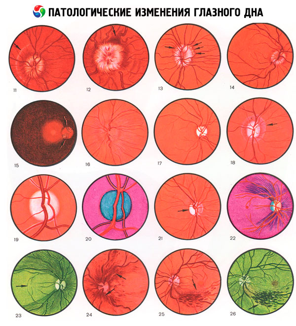
The method of ophthalmochromoscopy involves the use of certain light filters: red, yellow and blue, as well as polychromatic shades - the so-called red (blue-green), yellow-green and magenta.
Interpretation of the study is carried out taking into account the color transformation. For example, if you use a purple hue that passes only the rays of red and blue, any element that is not magenta-colored is separated. Often such information is useful: in particular, the pallor of the optic disc in the case of atrophy becomes bluish.
The yellow element under the blue light becomes almost black.
The yellow-green ray can be perfectly absorbed by the blood, and also reflected by the retina of the eye. As a result of this, hemorrhages, capillaries and even small aneurysms are clearly determined due to a strong increase in the contrast between the listed elements and the fundus.
Black elements on the background of yellow-green fabric are defined more clearly than the red elements on the reddish cloth.
Yellow-green rays increase the contrast and make the details more clear. This is due to the fact that the human eye is more sensitive to the yellow-green spectral shade.
All the available options for light filters have their pluses and minuses, so each of them uses a doctor for a specific purpose:
- red tint - helps to identify pigmented elements and defects in the shell of vessels;
- yellow tint - defines hemorrhages under the mesh skin of the eye, which have a dark brown color;
- blue tint - provides an opportunity to consider the mossy surface elements;
- purple hue - gives information on the severity of dystrophic changes in the retina;
- blue-green tint - indicates a matt opacity of the mesh shell, or rather, its central part.
Describing the results, the doctor lists all the detected pathological elements (if any), indicating their size, structure, parameters and depth of penetration. The characteristic changes in these elements in various spectra are noted without fail. In the final description, all results are combined with the results of other studies, on the basis of which the diagnosis is made or refined.
Ophthalmoscopy is performed by an ophthalmologist who will directly diagnose and prescribe the treatment. Such an approach excludes the appearance of inaccuracies and errors that can appear when information is transferred from one specialist to another.
Medical expert editor
Portnov Alexey Alexandrovich
Education: Kiev National Medical University. A.A. Bogomolets, Specialty - "General Medicine"