Medical expert of the article

New publications
Neuroretinitis
Last reviewed: 29.06.2025

All iLive content is medically reviewed or fact checked to ensure as much factual accuracy as possible.
We have strict sourcing guidelines and only link to reputable media sites, academic research institutions and, whenever possible, medically peer reviewed studies. Note that the numbers in parentheses ([1], [2], etc.) are clickable links to these studies.
If you feel that any of our content is inaccurate, out-of-date, or otherwise questionable, please select it and press Ctrl + Enter.
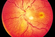
Neuroretinitis is more often a unilateral (less often bilateral) inflammatory process characterized by damage to the optic nerve and retinal nerve fiber layer, visual impairment, damage to the outer retina and retinal pigment epithelium. The exact origin of the disease is unknown, although it is accepted that intoxication is implicated in the development of inflammation. Neuroretinitis is a form of optic neuritis, characterized by a slowly progressive course and requiring long-term therapy. [1]
Epidemiology
Neuroretinitis is detected with an approximate frequency of 1 to 5 patients per hundred thousand population. Among all ophthalmologic diseases the pathology is registered in less than 3% of cases.
In many cases, neuroretinitis ends with restoration of visual function, but 25% of patients experience irreversible consequences in the form of loss or deterioration of vision. Some patients are disabled.
The disease affects men and women of all ages about equally. The average age of the diseased is 25-35 years. In most cases, neuroretinitis develops against the background of another infectious and inflammatory process in the body. [2]
Causes of the neuroretinitis
Cytomegalovirus neuroretinitis develops in patients with immune abnormalities - e.g. hIV. The inflammatory reaction develops in the area of the ocular fundus, further spreading to the retina. If the disease is not detected in time, there is a risk of retinal detachment in the future.
Syphilis neuroretinitis is a consequence of the third stage of syphilis, when the pathogen penetrates into the internal structure of the eye. Sometimes the pathology develops in infants: in this case, neuroretinitis is the result of hereditary pathology.
Toxoplasmosis can also be transmitted to the child in utero. Neuroretinitis is a consequence of this lesion and occurs in a person several years after birth.
Septic neuroretinitis is a complication of purulent-inflammatory processes in internal organs.
A viral lesion occurs as a result of a severe course of influenza, herpes and so on. In such a situation, most often a mild form of neuroretinitis develops, which passes as the underlying disease subsides.
Sometimes the causes are congenital vascular pathologies - for example, hemorrhagic retinitis (Coates disease, retinitis pigmentosa ). These diseases are caused by pathological changes in genes. [3]
Additional causes may include:
- Infection from other sites in the body;
- Trauma to the eye;
- Prolonged exposure to ionizing radiation;
- Regular exposure to ultraviolet light.
Risk factors
The exact factors in the development of neuroretinitis have not been determined. However, most often we are talking about infectious rhinogenic inflammatory processes, and inflammation can have different origins: bacterial, viral, fungal, parasitic, toxic. In general, any acute or chronic infectious disease can theoretically cause neuroretinitis.
In addition, pathology can develop as part of an autoimmune reaction - in particular, in patients with rheumatologic diseases. The problem is somewhat less often caused by traumatic damage to the organs of vision.
Additional factors:
- Age - the risks of neuroretinitis increase with age (the disease is more common in the elderly).
- Hereditary predisposition - some provoking pathologies are inherited.
- Harmful habits, poor diet, neuropathologies.
- Vascular disease, hypertension, atherosclerosis.
- Specific diseases (HIV, syphilis, etc.).
- Diabetes mellitus, pernicious anemia, ophthalmopathy.
Pathogenesis
Neuroretinitis is an inflammatory process involving the optic nerve and the retinal nerve fiber layer. The optic nerve is a segment of the peripheral neuron of the optic pathway. Its beginning is defined in the region of the eye fundus, and its completion - in the middle cranial fossa. It is formed by the axial cylinders of the retinal ganglia and is represented by approximately 1 million nerve fibers. The nerve exits the orbit through the optic orifice, after which both nerves are directed to the Turkish saddle. [4]
The development of neuroretinitis can be caused by both acute and chronic infection. Especially often the primary sources are otolaryngologic diseases (Maxillary sinusitis, sinusitis and tonsillitis ), dental pathologies (periodontitis or carious teeth), inflammations of the brain and brain membranes (Meningitis - serous, syphilitic or tuberculous, encephalitis - viral, rickettsial, bacterial or protozoal), as well as influenza, tuberculosis, syphilis, rye, etc.). [5]
Of the diseases of internal organs pathological sources are often kidney and blood diseases, allergic processes, diabetes mellitus, gout, collagenosis, avitaminosis. Intoxication - for example, alcohol-tobacco, lead, methanol - is also of considerable importance. A large percentage of neuroretinitis cases are of unexplained origin. [6]
Symptoms of the neuroretinitis
Cytomegalovirus neuroretinitis is characterized by signs such as:
- The appearance of small spots, flies in front of the eyes;
- The appearance of sparkling flashes (which is especially noticeable at night);
- Drop in visual acuity, formation of blind spots;
- Deterioration of peripheral visual function.
In syphilitic neuroretinitis, vitreous opacity, swelling of the retina and optic nerve are noted. Retinal hemorrhages are possible.
In septic complications, vitreous body opacity, optic nerve edema, and in advanced cases a pronounced purulent inflammation develops.
Neuroretinitis associated with pathological changes in genes is often accompanied by impaired color perception, blurring of the visible image, sharp narrowing of the visual field, and impaired spatial orientation.
In general, patients most often voice complaints about sharp deterioration of visual function, narrowing and loss of visual fields, impaired color perception (especially blue-green spectrum). Many patients experience light flashes and pain in the eyeball. [7]
Complications and consequences
Neuroretinitis can lead to visual impairment ranging from worsening to complete loss of visual function in either one eye or both eyes. Vision can deteriorate dramatically over several days. Sometimes 1-2 days are enough for the patient to lose more than 50% of visual function.
Color perception is particularly affected, but the patient may not notice or pay attention to this for a long time. Most patients with neuroretinitis experience intraocular pain, which increases during eyeball movements. In addition, the disease is prone to recurrence.
In the process of compressing or damaging the optic nerve axons, axoplasmic transport is disrupted. Optic nerve edema develops, the fiber is damaged, and the ability to see is impaired, which can cause partial or complete optic atrophy if treated incorrectly or late. [8]
Diagnostics of the neuroretinitis
The diagnosis of neuroretinitis is established on the basis of an ophthalmologic examination. At the first diagnostic stage, the doctor interviews the patient, analyzes the history of the disease, clarifies the results of examination of other specialists (neurologist, endocrinologist, neurosurgeon), performs a complete ophthalmologic examination and assesses the probability of possible symptomatology of various neuropathologies. If necessary, prescribes a number of additional examinations and determines further treatment regimen.
Mandatory tests for the diagnosis of neuroretinitis:
- General blood examination (to exclude chronic inflammation and systemic autoimmune process);
- Urinalysis;
- Biochemical blood test with determination of glucose, AST, ALT;
- Bacteriologic seeding from the conjunctival cavity with determination of the causative agent and its sensitivity to antibiotic therapy;
- Blood tests for syphilis (RW) and HIV by ELISA;
- ELISA analysis of hepatitis B and C markers;
- Ig A, M, G analysis to herpes simplex, chlamydia, cytomegalovirus, toxoplasmosis viruses.
Additional recommendations may include:
- C-reactive protein blood test;
- Blood test for rheumatic tests.
Instrumental diagnosis is often represented by basic diagnostic procedures such as:
- Visometry is a traditional method of assessing visual acuity;
- Biomicroscopy - a technique for detecting lens opacity, focal or diffuse vitreous opacity, vitreous hemorrhages, cells, exudate, hypopyon;
- Tonometry is a method of determining intraocular pressure;
- Ophthalmoscopy - investigation of changes in the posterior ocular segment, inflammatory foci, muffs along the vessels, intraretinal hemorrhages, hard deposits, macular edema, neuropathy, atrophic changes of the optic nerve characteristic of chorioretinal inflammation;
- Perimetry - assessment of possible narrowing of the visual field, detection of scotomas, diagnosis of central and peripheral vision dysfunctions;
- Refractometry - detection of ocular refractive disorders;
- X-ray of the sinuses and chest - to exclude pathologic processes that could cause the development of neuroretinitis.
Eye fundus biomicroscopy, gonioscopy, examination of the periphery of the eye fundus, ophthalmochromoscopy, electroretinogram, ultrasound examination of the eyeball and cerebral vessels, optical coherence retinotomography, fluorescence angiography, X-ray of the orbit and skull in different projections may be prescribed if indicated.
Registration of evoked visual potentials is often used, which is necessary to assess the state of the optic nerve and differential diagnosis of neuroretinitis from functional and organic visual disorders. [9]
Differential diagnosis
|
Pathology |
Basis for differential diagnosis of neuroretinitis |
|
Secondary central chorioretinal dystrophic process |
There is evidence of past ocular inflammation. There is a central scotoma in the visual field. |
|
Age-related degenerative process in the macula |
There is a central scotoma in the visual field, a drop in visual acuity is noted. |
|
Retinitis pigmentosa |
There are defects in the visual field, a drop in visual acuity. Ophthalmoscopy reveals various pathologic foci in the retinal area. |
|
Chorioid tumors |
There is a drop in visual acuity, and ophthalmoscopy reveals a focal area with indistinct outlines, indentation. |
|
Chorioretinopathy, central serous in nature |
There is a sharp deterioration of vision, sometimes associated with a viral illness. |
|
Epitheliopathy, acute placoid multifocal type |
Vision decreases after a viral illness, paracentral or central scotomas are noted. Photopsia, metamorphopsia may be detected. |
|
Subretinal and subchoroidal hemorrhages |
Vision decreases sharply, scotoma appears in the visual field. Ophthalmoscopy reveals a focus with indistinct outlines. |
|
Hemorrhagic retinal detachment |
Vision decreases sharply, scotoma appears in the visual field. Ophthalmoscopy reveals a pathologic focus in the retinal area. |
Who to contact?
Treatment of the neuroretinitis
Conservative therapy may include various medications, which depends on the cause of neuroretinitis.
If pupil dilation is necessary, cycloplegic and mydriatic drugs are prescribed:
- 1% tropicamide - 2 drops twice a day, for a week;
- 1% phenylephrine 2 drops twice daily for a week.
Glucocorticosteroids are used to block the inflammatory response in neuroretinitis, reduce capillary permeability, inhibit the production of prostaglandins, slowing down proliferation processes:
- 0.1% dexamethasone 2 drops. 4-5 times a day;
- 0.4% dexamethasone once daily 1.2-2 mg under the conjunctiva or 2-2.8 mg parabulbarly;
- Prednisolone 5 at 30-80 mg daily orally (in the morning) with further gradual reduction of the dose for 10 days (indicated in regularly recurrent neuroretinitis, systemic pathologies);
- Methylprednisolone 250-1000 mg daily intravenous drip for 4-5 days (if local treatment is ineffective, or there is a severe chorioretinal inflammation with an increasing threat of loss of visual function, or in bilateral neuroretinitis associated with systemic pathologies).
In neuroretinitis due to infectious processes, antibiotic therapy is indicated:
- 0.3% Tobramycin 2 drops. 5 times a day;
- 0.3% Ciprofloxacin 2 drops. 5 times a day for a week;
- Levofloxacin or Moxifloxacin 2 drops. 5 times a day for a week;
- Ciprofloxacin 250-500 mg daily orally for a week;
- Amoxicillin 250-500 mg daily orally for two weeks;
- Clindamycin 150 mg orally 4 times a day for 1-2 weeks;
- Ceftriaxone 1 g daily as intramuscular injections, a course of 1-2 weeks;
- 30% Lincomycin 600 mg twice daily as intramuscular injections, course of 1 week.
If neuroretinitis is provoked by a viral disease, antiviral therapy is prescribed:
- Acyclovir 200 mg 5 times a day for a week;
- Valacyclovir 500 mg three times a day for a week.
If neuroretinitis is caused by a fungal pathogen, antifungal therapy is appropriate:
- Ketoconazole 200 mg twice a day orally, for 1-2 weeks;
- Fluconazole 150 mg twice daily for 10 days.
When neuroretinitis is combined with increased intraocular pressure, diuretics are prescribed:
- Furosemide 40 mg daily for three consecutive days;
- Furosemide 1% by 2 ml as intramuscular injections daily for 2-3 days.
Nonsteroidal anti-inflammatory drugs are indicated to block the inflammatory response:
- Diclofenac sodium 25-75 mg daily intramuscularly for a course of 5 days;
- Meloxicam 15 mg daily as intramuscular injections for a course of 5 days;
- Indomethacin 25 mg three times a day orally for 2 weeks.
In complicated cases of neuroretinitis, systemic and frequently recurrent pathologies, absence of positive response from glucocorticosteroids it is possible to prescribe antimetabolites (Methotrexate, 5-fluorouracil in the subtenon space). [10]
The effectiveness of the treatment is evaluated by the following indicators:
- Improved vision;
- Eliminating the inflammatory response;
- Resorption of the infiltrate;
- Decreased severity of object distortion, photopsia, scotoma.
Surgery is not indicated for neuroretinitis.
Prevention
Preventive measures should be carried out for all people who have a tendency to develop neuroretinitis (including genetic predisposition to pathology):
- Have regular check-ups and consultations with eye specialists;
- Avoid head and eye injuries;
- Do not self-medicate for any infectious diseases (including the common cold);
- Keep physically active, avoid hypodynamia;
- Give up bad habits;
- Eat a varied, balanced diet;
- Do not overwork your eyes, avoid spending long periods of time in front of a computer screen or gadgets;
- Enough rest, getting at least 7-8 hours of sleep a night;
- Have regular blood and urine tests to assess performance;
- Take frequent walks in the fresh air;
- Avoid activities involving excessive visual strain;
- Regularly visit the dentist, prevent the development of dental caries, periodontitis.
In addition, to prevent neuroretinitis, it is recommended to use sunglasses to protect the retina from ultraviolet light, periodically check with specialists to eliminate risk factors.
Forecast
The prognosis depends mainly on the underlying cause of neuroretinitis - that is, on the course of the underlying pathology. Some mild inflammatory processes resolve on their own, and vision returns in a few weeks (months). In the absence of dynamically unstable and systemic diseases (connective tissue pathologies), visual function can be restored, but often the problem becomes recurrent, affecting the same or another eye.
To optimize the prognosis, it is necessary to timely treat acute and recurrent pathological processes, eliminate bad habits, regularly visit specialized specialists and conduct preventive examinations. [11]
If neuroretinitis progresses to a chronic form, the risk of complications and adverse effects increases dramatically.