Medical expert of the article

New publications
Myxoma of the heart and soft tissue
Last reviewed: 04.07.2025

All iLive content is medically reviewed or fact checked to ensure as much factual accuracy as possible.
We have strict sourcing guidelines and only link to reputable media sites, academic research institutions and, whenever possible, medically peer reviewed studies. Note that the numbers in parentheses ([1], [2], etc.) are clickable links to these studies.
If you feel that any of our content is inaccurate, out-of-date, or otherwise questionable, please select it and press Ctrl + Enter.
For a heterogeneous group of primary neoplasms of soft tissues in the form of benign mesenchymal tumors, there is a definition such as myxoma.
This term was introduced in the second half of the 19th century by the famous German pathologist Rudolf Virchow.
 [ 1 ]
[ 1 ]
Epidemiology
Since this type of tumor is a rare pathology, general statistics are not kept, and the WHO only registers the prevalence of cardiac myxomas – at a level of 0.01-0.02%. In approximately 5% of cases, myxoma is an inherited sign of a family genetic pathology.
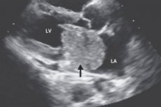
Myxomas account for 48% of primary benign cardiac tumors in adults and 15% in children. Most tumors, according to the European Journal of Cardio-Thoracic Surgery, are localized in the left atrium (60-87%).
The right ventricle accounts for 8% of myxomas, while left ventricular myxoma is diagnosed in approximately 4% of cases. The mitral valve accounts for 6% of myxoma cases, and multiple tumors are diagnosed in 20% of patients.
Myxomas are three times more common in women; the average age range of patients is 44-56 years.
According to some data, intramuscular myxoma mostly affects people after 50, and the incidence rate is 0.1-0.13 per 100 thousand people.
Causes myxomas
Such neoplasms are rarely detected, and to date, the specific causes of myxoma are unknown. It is not precisely determined how long myxoma grows, but these sporadic tumors are characterized by a long period of formation. By the way, they are found not only in soft tissues of almost any localization, but also in the area of joints.
The key histological feature of myxomas is the presence of a mucous (mucopolysaccharide) mass, often encapsulated, with fibroblast cells freely embedded in it, which makes it similar to mesenchyme - the tissue from which all connective tissues, blood vessels and muscle fibers are formed during the antenatal development of the body.
Typically, myxomas have an oval or spherical shape, a gelatinous surface, and a fibrous capsule that grows into adjacent muscle tissue on a thin stalk or wide base.
[ 6 ]
Pathogenesis
Apparently, the pathogenesis is caused by a violation of the differentiation of mesenchymal cells and the formation of modified fibroblasts that produce excess sulfated mucopolysaccharides (glycosaminoglycans) and immature fibrous tissue cells.
In an attempt to determine the etiology of this type of neoplasm, researchers have found that about 7% of all myxomas that form in cardiac structures are associated with the hereditary Carney complex, which includes – in addition to myxomas of the heart and skin – hyperpigmentation of the skin, primary nodular adrenocortical dysplasia (which manifests itself as symptoms of hypercortisolism) and pituitary adenoma with increased secretion of somatotropic hormone.
This syndrome is caused by a deletion of the gene encoding the protein kinase A enzyme in locus 17q2, which plays a leading role in the process of differentiation of structural proteins, as well as the growth and division of cells of all tissues of the body. Moreover, as foreign clinical practice shows, in 8 cases out of 10, patients with this genetic pathology first develop a skin myxoma, and after a while, a myxoma forms in the heart.
In addition, it is revealed that aberrations in chromosomes 2, 12, 13 and 15 are involved in the formation of cardiac myxoma. However, no more than 10-12% of myxomas are considered genetic; in other cases, these tumors are recognized as idiopathic.
Symptoms myxomas
Depending on the location of the tumor, both the first signs of myxoma development and its clinical symptoms at later stages differ.
For example, when a tumor grows in internal organs or skeletal muscles, its only symptom is the presence of a growing homogeneous mass that does not cause pain or inflammation and is often discovered by chance.
In the initial stage, cardiac myxomas do not manifest themselves in any way, and in about 15% of cases they are completely asymptomatic. But as the tumor grows, heart failure may develop - with shortness of breath during exertion (up to orthopnea), attacks of difficulty breathing at night (due to pulmonary edema), ascites and hepatomegaly. Patients complain of arrhythmia and chest pain, note persistent cyanosis of the skin and especially of the fingers (indicating problems with blood circulation).
Left atrial myxoma can cause atrioventricular valve insufficiency due to the constant movement of the tumor mass, which prevents them from closing and can damage the tendon threads (chords) in the heart. Symptoms of myxoma in this location are very similar to those of mitral stenosis and congestive heart failure, including dizziness, breathing problems, coughing and hemoptysis, severe cachexia with subfebrile temperature.
Myxoma of the right atrium can cause symptoms of pulmonary hypertension: increased fatigue, severe shortness of breath during the day, peripheral edema of the lower extremities, fainting, cough.
A large right ventricular myxoma manifests itself with symptoms of pulmonary artery narrowing in the form of angina pain, dyspnea, and fainting. A left ventricular myxoma at the progression stage causes a hemodynamic disorder such as a ventricular filling defect in half of the patients – due to impaired blood flow through the mitral valve.
When a periarticular myxoma forms, joint pain and decreased mobility are possible. Cutaneous myxomas occur in patients with Carney syndrome and are single or multiple encapsulated concretions – soft nodules (up to 2.5 cm in diameter) of flesh color, often with blood vessels – on the face, trunk or limbs.
Myxoma of the abdominal cavity, which has a collagen or fibrous membrane, when reaching significant sizes sometimes causes a feeling of discomfort and dull pain. And with myxoma of the vermiform appendix, there may be the same signs as with chronic inflammation of the appendix.
Forms
In the international classification of soft tissue tumors – WHO Classification of Soft Tissue Tumors (4th edition 2013) – among all classes of benign neoplasms, the definition of “myxoma” is found in the class of tumors of uncertain differentiation (G9).
Experts have identified the following types: intramuscular myxoma, periarticular myxoma, superficial angiomyxoma, deep (locally aggressive) angiomyxoma, dermal myxoma of the nerve sheaths (neurothecoma).
Cardiac myxoma is not distinguished in this classification, but cardiologists note the following varieties: atrial myxomas - left atrium (usually detected after 40 years) or right atrium (localized on the atrial septum); ventricular myxomas (formed in the ventricles of the heart), mitral valve (extremely rare).
Intramuscular myxoma is formed in the depth of the skeletal muscles of the upper and lower extremities - myxoma of the thigh, myxoma of the leg; in the muscle tissue of the shoulders or buttocks. The tumor can occur in isolation, as well as in combination with Albright syndrome. Multiple formations in muscle tissue against the background of fibrous dysplasia (replacement of bone tissue with fibrous) are defined as Mazabroud syndrome.
Periarticular myxoma can be found in the shoulder or elbow; in the knee area (88% of cases), hip joint, ankle or heel. Doctors note risk factors for the appearance of such formations: osteoarthritis of the joint or previous injuries.
Locally invasive types include myxoma of the jaw, a rare intraosseous neoplasm that most often occurs on the lower jaw. It is classified as a slowly growing odontogenic tumor, i.e. formed from the mesenchymal part of the tooth germ. Myxoma is possible on the palate in the mouth, on the gum or cheek.
With the help of hardware visualization, tumors of this group can be identified at the base of the skull and on the temporal bone, in the neck area, as well as left-sided supraclavicular (periclavicular) formation or myxoma of the right supraclavicular region.
In adults, along with serous and mucinous cysts or pelvic arteriovenous malformations, CT and MRI reveal such a primary pelvic retroperitoneal neoplasm as pelvic myxoma or hip retroperitoneal myxoma.
Benign tumors that form in the retroperitoneal space: extra-organ aggressive angiomyxoma or myxoma of the abdominal cavity, as well as of the vermiform appendix (appendix), which is more often diagnosed as a mucocele (mucous cyst) of the appendix, which may be associated with peritoneal pseudomyxoma (histologically representing a mucinous adenocarcinoma or cystadenoma).
Also, aggressive (infiltrative) angiomyxoma can be anogenital – myxoma of the labia, vulvovaginal area and perineum, and its appearance is most likely in patients at menopause age.
Complications and consequences
Although these tumors are benign in nature, they have serious consequences and complications.
As cardiologists note, the most dangerous complications of cardiac myxoma are systemic embolism, which occurs in 30-45% of patients with left atrium tumors and in 10% of cases of right atrium tumors. Left ventricular myxomas have the highest embolism rate (more than 60%).
Embolism develops due to the separation of tumor fragments and their entry into the bloodstream, which can result in blockage of the coronary arteries with the development of a heart attack, an increase in pressure in the pulmonary circulation (development of pulmonary hypertension), and the cessation of blood flow in the pulmonary artery.
Emboli can affect cerebral vessels, causing cerebral infarction and neurological damage: visual impairment, seizures, hemiparesis, aphasia and progressive dementia.
Large atrial fibroids can cause narrowing of the heart valves—mitral or tricuspid stenosis—and sudden death.
Sequelae associated with Carney complex include recurrent growth of myxomas in approximately 12-22% of familial cases.
An odontogenic tumor of the upper jaw can lead not only to facial deformation, but also to difficulty breathing or obstruction of the maxillary sinus.
[ 15 ]
Diagnostics myxomas
The correct diagnosis of myxoma requires a lot of clinical experience, and for each type of these formations there are differences in diagnostic procedures. Myxomas of the skin require histology; to identify the Carti complex, it is necessary to analyze some immunohistological markers (the analysis is carried out after the tumor is removed).
Blood tests are required: general, electrolyte and troponin levels, β-globulin (blood clotting factor VIII), ESR, C-reactive protein, immunoglobulins (IgM, IgE and IgA), thyroid hormone levels and ACTH.
To date, markers for myxoma localized to the heart include interleukin-6 (IL-6) and interleukin-8 (IL-8) in serum, as well as phospholipase A2.
The results of the studies showed that monoclonal antibodies to the transmembrane protein CD34, associated with the differentiation of prolonged hematopoietic stem cells (LT-HSC), can be a marker of this type of neoplasm in other structures of the body.
Instrumental diagnostics of cardiac myxoma uses ECG, transesophageal and transthoracic ultrasound echocardiography, angiocardiography, and magnetic resonance imaging.
Differential diagnosis
Very important differential diagnosis of myxomas. Thus, differential diagnosis of cardiac myxoma includes distinguishing its symptoms from signs of heart defect, cardiomegaly, bacterial endocarditis, primary pulmonary hypertension, pulmonary embolism, regurgitation and/or stenosis of the mitral/tricuspid valve, as well as from fibrosarcoma, lipoma, hemangioma, desmoid tumor.
Myxoma within muscle tissue may be mistaken for sarcoma. And cutaneous myxomas should be differentiated from lipomas, dermatofibromas, intraepithelial cystic lesions, basal cell epithelioma, or basal cell carcinoma.
Ultrasound, CT and MRI are used to visualize the masses of other locations. Doctors detect the tumor but cannot differentiate it, so an accurate diagnosis can only be made after surgical removal of the formation and its histological examination.
Who to contact?
Treatment myxomas
Surgical treatment, that is, complete removal of a symptomatic myxoma, is recognized as the only correct method of treating these neoplasms.
It is believed that only surgery for cardiac myxoma – sometimes urgent due to the severity of symptoms and in all cases requiring the patient to be connected to an artificial blood circulation machine (ABM) and the use of hypothermic cardioplegia – can prevent the development of life-threatening complications.
The list of absolute contraindications to immediate surgical intervention includes stroke and cerebral hemorrhage.
Access to the tumor, the technique of its removal, as well as the need for manipulations on the heart valves (their annuloplasty or replacement with an endoprosthesis) are determined depending on the localization of the myxoma. But a mandatory condition is extensive resection of all tumor tissues and excision of the site of its attachment to exclude relapse, and minimal impact on the tumor - to avoid local embolism during surgery.
Rehabilitation after myxoma removal varies from patient to patient: it all depends on the complexity of the operation and the condition of the individual patient. But in any case, it is a fairly lengthy process.
According to European clinics, the mortality rate after such operations is 5-6%.
[ 20 ]