Medical expert of the article

New publications
MRI of the ocular orbits
Last reviewed: 04.07.2025

All iLive content is medically reviewed or fact checked to ensure as much factual accuracy as possible.
We have strict sourcing guidelines and only link to reputable media sites, academic research institutions and, whenever possible, medically peer reviewed studies. Note that the numbers in parentheses ([1], [2], etc.) are clickable links to these studies.
If you feel that any of our content is inaccurate, out-of-date, or otherwise questionable, please select it and press Ctrl + Enter.
Magnetic resonance imaging is a method of diagnostic examination of various human organs, combining knowledge of nuclear physics and medicine. This method is a little less than 60 years old, but it began to be actively used only at the turn of the last and current centuries directly for the study of internal organs and the brain. A little later, the method gained great popularity in ophthalmology for the diagnosis of eye diseases, the cause of which is not visible during a visual examination. MRI of the orbits and optic nerves allows you to identify the slightest changes in various tissues and structures of the eye that affect a person's ability to see. This means that this method helps to identify the disease at its initial stage and begin treatment when it will be most effective.
 [
[ Indications for the procedure
Magnetic resonance imaging is considered one of the safest and most effective ways to detect various eye pathologies by carefully examining internal structures that are not visible to the naked eye and are not visible during examination with a microscope. In addition, the more modern MRI method helps to see such minor changes in the eye that are inaccessible to study using old methods.
Due to the high diagnostic value of orbital MRI, it can be prescribed for diagnosing a wide variety of eye pathologies:
- inflammatory processes localized in various layers of the visual organ,
- damage to the retina, such as retinal detachment,
- tumor processes in the organ area with determination of their exact location and size (even small neoplasms from 1 mm are determined),
- hemorrhages in the eye with determination of their cause, thrombosis of the vessels of the eye,
- injuries with determination of the severity and volume of damaged tissues, with identification of remnants of foreign bodies that caused eye injury,
- changes in the corneal layer,
- dysfunction of the optic nerves (for example, if glaucoma is suspected ), decreased visual acuity, the appearance of unexplained pain in the eye with determination of its cause,
- the condition of the organ of vision in diabetes mellitus, hypertension and other pathologies in which the blood supply to the eye is disrupted.
MRI can be used to determine the location of foreign bodies in the internal structures of the eye, identify inflammatory foci and assess their size, find hidden tumors and take biopsy material under MRI control.
If there has been an eye injury, MRI allows us to assess its consequences and complications, the size and nature of damage to internal structures as a result of the injury, and the possibilities of treatment in each specific case.
When a person's vision deteriorates or the motor activity of the eyes is impaired ( strabismus appears, the patient cannot focus his vision on a certain object), it is simply impossible to determine the cause without examining the internal structures. MRI makes it possible to see and assess the degree of damage (atrophy) of the muscles or nerves responsible for eye movement, and to outline measures to correct the defect.
Quite often the cause of visual impairment and pain is hidden from us, and it can be detected only by virtually penetrating inside the eye, observing its work, assessing the changes occurring there. This is the opportunity that magnetic resonance imaging provides. And although the procedure is called MRI of the orbits, in fact, it also allows visualizing disorders of the visual muscles, nerves and lacrimal glands, pathologies of the eyeball, changes in fatty tissue, due to which its demand is growing.
Preparation
MRI of the orbits and optic nerves is considered a simple and generally safe procedure that does not require special measures to prepare for diagnostics. It is usually prescribed by an ophthalmologist during an appointment and examination of the patient if the patient has difficulty making an accurate diagnosis.
A person can undergo an examination on the same day or later, when such an opportunity arises. The fact is that not all medical institutions are equipped with the necessary equipment. In addition, the MRI procedure will not be free for everyone.
The main condition for obtaining a high-quality image is the patient's immobility during the examination, which the person is warned about in advance. If the patient is very nervous, has symptoms of claustrophobia or severe pain that does not allow him to remain still, sedatives are indicated to reduce nervous excitability.
Patients with mental disorders or serious eye injuries that cause unbearable pain require additional immobilization of the limbs. If the above measures do not help, the doctor may resort to anesthesia administered intravenously.
Since the examination of organs is carried out using a magnetic field, any metal objects that can distort it must be removed. This includes jewelry and clothing with metal elements (locks, buckles, buttons, decorative overlays, etc.). If there is metal in the body in the form of crowns, organ implants, electronic devices that support body functions, you need to tell the doctor about it during the appointment. It may be necessary to clarify the material of dentures if the patient is not sure of his information.
During MRI, contrast agents can be used, which facilitate the diagnosis of tumor and inflammatory processes, help to assess the condition of blood vessels. This issue is also discussed in advance, because the day before the procedure (5 hours before it) the patient will have to abstain from food, so that no food components can affect the results of the study. The optimal option is considered to be the introduction of contrast on an empty stomach.
To exclude intolerance to the contrast agent and anaphylactic reactions, a test is performed before the drug is administered, applying the drug to open areas of skin in the wrist area. The doctor must specify the patient's weight, because the volume of the contrast administered depends on it.
The drug is administered intravenously as injections or infusions (drip) into the elbow area. The patient may feel dizziness, heat, hot flashes, nausea, but this is not scary, since it is considered a normal reaction of the body to contrasts. The introduction of drugs for MRI of the orbits with contrast is carried out under the supervision of a doctor. For the next 30 minutes, the patient is monitored by medical personnel.
Half an hour after the administration of the drugs, the active substance of which accumulates in different tissues in different concentrations, you can begin MRI diagnostics. During this time, the drug will spread through the bloodstream and reach the area being examined.
Technique MRI of the ocular orbits
Orbital MRI, like any other diagnostic procedure, is not done for the sake of interest. Therefore, it should be taken seriously. After examining the patient, the specialist gives a referral for a diagnostic examination. With this referral and the results of previous examinations of the visual organs, the patient is sent to the diagnostic room.
The X-ray we are used to is somewhat different from magnetic resonance imaging, although both studies are identical and pursue the same goals. An uninitiated person may be a little shocked by the device in the form of a long, volumetric tube located horizontally. It is in this tube (capsule) that a magnetic field is created, allowing an image of the organ being examined to be obtained on the screen in all details.
To relieve tension and fear of the device and the procedure, the patient is explained how an MRI of the eye is performed, what the procedure can show in each specific case, and what consequences this study has for the body.
The operating principle of magnetic resonance installations of open or closed type is based on the recording of the movement of hydrogen atoms saturating the tissues of the body under the influence of a magnetic field. The illumination of different areas of the image depends on the number of gas molecules accumulated there.
The MRI procedure is quite complex to perform and requires the patient to remain still. This is easiest to do in a horizontal position, when the person is as relaxed as possible. For these purposes, the tomograph has a sliding table on which the patient is placed, fixing his head in a special device. If necessary, other parts of the body can be fixed with belts.
Since only the head area is being examined, the table is shifted so that only the head is inside the machine. The torso is outside the tomograph.
Before the procedure, patients are asked to use earplugs, as the device produces an unpleasant monotonous sound that can cause anxiety and unwanted movements.
The procedure itself is considered to be quite lengthy compared to X-rays. It takes from 20 to 40 minutes, during which the person must lie still. If contrast agents are used during the examination, the procedure may take another twenty minutes.
During the examination, the doctor is usually outside the diagnostic room, but the patient can contact him/her via speakerphone at any time if there is an attack of claustrophobia or any other problem, such as chest pain, shortness of breath, or a feeling of lack of air, which happens during the procedure with contrast. In the same way, the doctor can give the necessary instructions to the patient.
To reduce nervous tension and calm the patient, it is allowed to invite relatives to the procedure. This is especially important if the diagnostics are performed on a child. After all, the MRI machine is universal, so it is large and can frighten a small patient.
Contraindications to the procedure
Magnetic resonance imaging (MRI) is considered one of the safest procedures, because unlike computed tomography (CT) and radiography, it does not require the use of harmful X-rays. The magnetic field in the tomograph does not harm the health of a person of any age and condition, so health problems are more likely to be indications for the study than contraindications to it.
The only absolute contraindication to MRI is the presence of ferromagnetic alloys and electronic devices (pacemakers, electronic middle ear implants, etc.) in the human body. The magnetic field can negatively affect the operation of the pacemaker, simulating the heart rhythm and cause failures in the operation of electronic microscopic equipment implanted in the body.
As for metal implants made of ferromagnetic alloys and metal fragments stuck in the body (for example, after injuries), the danger of the influence of a strong magnetic field is that under its influence ferromagnets can noticeably heat up, causing tissue burns, and move from their place. Thus, the magnetic field can negatively affect ferromagnetic and large metal implants, Elizarov devices, ferromagnetic simulators of the middle ear, prostheses of the inner ear containing ferromagnetic elements, vascular clips made of ferromagnets installed in the brain area.
Some metal implants (insulin pumps, nerve stimulators, valve prostheses, hemostatic clips, dentures, braces, endoprostheses, etc.) can be made of materials with weak ferromagnetic properties. Such implants are classified as relative contraindications, but they must be reported to the doctor, indicating the materials from which the device is made. After all, even these devices may contain ferromagnetic elements, and the doctor must assess how dangerous the effect of a magnetic field on them will be.
As for dentures, most of them are made of titanium, a metal with weak ferromagnetic properties, i.e. the magnetic field during MRI is unlikely to cause a reaction from the metal. However, titanium compounds (for example, titanium dioxide, used in tattoo paints) may react differently to a strong magnetic field, causing burns on the body.
In addition to non-ferromagnetic implants, relative contraindications include:
- early pregnancy (there is not enough information about the effect of magnetic fields on fetal development during this period, but this method is considered more preferable and safer than CT or X-ray),
- heart failure in the stage of decompensation, serious condition of the patient, need for constant monitoring of the body's functioning, bronchial asthma, severe dehydration
- fear of enclosed spaces or claustrophobia (due to the impossibility of conducting research on a person who, due to fear, cannot remain still for half an hour or more),
- inadequate condition of the patient (alcohol or drug intoxication, mental disorders will not allow clear images to be taken due to constant motor reactions),
- tattoos on the body made using paints containing metal particles (there is a risk of tissue burns if these are ferromagnetic particles).
- inner ear prostheses that do not contain ferromagnetic materials.
In these cases, the decision on the possibility of performing an MRI of the orbits is made by the doctor, taking into account the possible negative impact. In some cases, it is advisable to postpone the procedure for the time necessary for the patient's condition to normalize.
If we are talking about MRI with contrast, the list of contraindications becomes longer, since it requires the introduction of chemicals into the body, the reaction to which can be dangerous.
MRI with contrast is not performed:
- pregnant women regardless of the gestational age due to the ease of penetration of drugs through the placental barrier (the effect of contrast agents on the fetus has not yet been studied),
- in chronic renal failure (the contrast is eliminated from the body within 1.5-2 days, but in case of renal dysfunction it may be retained for a longer period, since the recommended consumption of large amounts of liquid is considered unacceptable),
- in case of hypersensitivity to contrast agents due to the risk of developing severe allergic and anaphylactic reactions.
- patients with hemolytic anemia.
Before undergoing an MRI procedure, for his own benefit, the patient is obliged to tell about any metal objects in his body, including fragments from wounds, tattoos and cosmetics used (and it is better not to use cosmetics), remove all types of jewelry, watches, clothing with metal elements.
Normal performance
MRI of the orbits and optic nerves is a diagnostic examination that is prescribed for a specific purpose. The purpose of the examination is to identify pathological processes in the eye tissues or to evaluate the results of treatment if MRI is prescribed again.
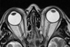
MRI allows for a detailed study of the shape and quality of development of the eye sockets, the location and shape of the eyeballs, the condition of the fundus, the structure and course of the optic nerve, and the identification of dystrophic changes in it and other abnormalities.
Using MRI of the orbits, it is possible to assess the condition of the ocular veins and muscles responsible for the movements of the eyeball (their location, the presence of seals and tumors), and the fatty tissue of the eye sockets.
MRI is used to detect damage to the retina, which is the inner lining of the eye. The fact is that damage to the retina does not necessarily have to be associated with eye or head trauma. Some pathologies of the inner lining of the organ of vision are associated with various systemic diseases (diabetes mellitus, hypertension, kidney and adrenal pathologies). Magnetic resonance imaging helps to detect such pathologies as retinal detachment, diabetic or hypertensive retinopathy, damage to the vessels that supply nutrition to the retina, dystrophy or degeneration of this part of the eyeball, tumor and inflammatory processes, retinal rupture.
MRI of the orbits with contrast allows you to assess the condition of the eye vessels, their blood filling, the presence of blood clots and ruptures. With the help of contrast agents, it is easier to recognize internal inflammations. But most often, the technique is still used to detect tumors when oncology is suspected. With the help of MRI, you can not only detect a tumor in a certain area of the eye, but also assess its shape and size, the presence of metastases, the effect on nearby structures and the possibility of removal.
Any deviations in shape, size, tissue density, detected by MRI of the orbits, provide the doctor with valuable information necessary for making a final diagnosis. In addition, during diagnostic procedures, it is possible to detect some damage to the brain, which is also visible on the tomogram.
An example of an orbital MRI protocol might look like this:
Type of study: primary (if the study is repeated, the date of the previous one is also indicated, with which the results will be compared).
The eye sockets are correctly developed, pyramidal in shape with clear and even contours of the walls. There are no foci of destruction or compaction.
The eyeballs are spherical in shape and symmetrically located relative to the eye sockets. The vitreous tissues are uniform, no changes in the MR signal are observed (this indicates a normal state of the organ, for example, in inflammatory processes the MR signal will be hyperintense, in tumors - isointense or hyperintense).
There is no thickening of the eye membranes. They have smooth and clear contours.
The optic nerves are characterized by a regular course and clear contours without dystrophic changes or local thickenings.
Orbital structures: The muscles of the eyeball are correctly positioned, there are no thickenings on them. Fatty tissue, ocular vessels and lacrimal glands are normal. The grooves of the convexital surface of the brain are unchanged.
Visible structures of the brain: There is no displacement of the midline structures. The cisterns of the brain base are not deformed. The lateral ventricles of the brain are of normal size and symmetrical location. There are no areas of pathological density in the area of the brain structures.
Other findings: none.
The MRI protocol (decoding) described above indicates that no pathological changes in the human visual organs were detected.
After receiving the image and the examination protocol (and you will have to wait about 30 minutes for them), the patient is sent to see an ophthalmologist, and sometimes a neurologist, to make a final diagnosis and prescribe the necessary treatment.
Complications after the procedure
Magnetic resonance imaging is one of the safest examinations, allowing you to scan various organs without harm to health, and also to obtain a three-dimensional image for a more detailed examination of the diagnostic object. Even though the eyes and brain are considered the most sensitive parts of the body, too susceptible to the influence of various negative factors, MRI is performed without fear for the health of these organs, since it does not carry a radiation load on these important, but very delicate structures. The magnetic field used in modern tomographs does not cause any consequences for the eyes and vital organs.
MRI of the orbits is a non-invasive procedure, i.e. it is possible to examine the internal structures of the eye without opening the tissues. This is another advantage of the modern diagnostic method.
Under MRI control, additional diagnostic studies can be performed, for example, a biopsy if a malignant tumor process inside the eye is suspected. And the tumor can be easily detected at an early stage of its development at a small size. This is ideally helped by MRI with contrast.
A three-dimensional image allows you to assess the condition of the organ in detail, the only thing is that it is not possible to obtain a clear image of the walls of the eye sockets, but all other structures are determined with great accuracy and without the health hazard that exists when performing CT. The safety of the magnetic resonance method allows it to be used in the diagnosis of ophthalmological and other diseases in children. However, the procedure is prescribed to children over 7 years old who are already able to remain motionless for a long time and follow the doctor's instructions.
The disadvantages of the method include high cost, relatively long duration of the procedure with the need to maintain a static position during the entire examination period (which is not as easy as it seems), the likelihood of heart rhythm disturbances and a large number of contraindications associated with metal and electronic implants.
However, safety for the body is more important than any money, and time is not an issue when it comes to accurate diagnostics and human health. Those categories of people who cannot undergo an MRI examination can resort to other diagnostic methods (X-ray, slit lamp, biomicroscopy of the eye, etc.), so they will not be left without the help of doctors.
Complications during MRI of the orbits can only occur if contraindications to the procedure are ignored. And in most cases, they are limited to minor tissue burns or distortion of the results of the study, if the patient does not report a tattoo or implant. Usually, those people who have devices installed that monitor the functioning of vital organs and systems do not forget about them and always report them before prescribing diagnostic tests. But if the information was hidden intentionally, this is the responsibility of the patient himself, who was informed about the requirements for high-quality diagnostics before the procedure.