Medical expert of the article

New publications
Magnetoencephalography
Last reviewed: 06.07.2025

All iLive content is medically reviewed or fact checked to ensure as much factual accuracy as possible.
We have strict sourcing guidelines and only link to reputable media sites, academic research institutions and, whenever possible, medically peer reviewed studies. Note that the numbers in parentheses ([1], [2], etc.) are clickable links to these studies.
If you feel that any of our content is inaccurate, out-of-date, or otherwise questionable, please select it and press Ctrl + Enter.
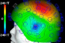
Magnetoencephalography is the registration of the magnetic component of the electromagnetic field of the brain. This method arose relatively recently due to the successes of low-temperature physics and ultra-sensitive magnetometry.
Magnetoencephalography is not only a non-invasive, but even a contactless method of studying the functional state of the brain. Its physical essence lies in the registration of ultra-weak magnetic fields that arise as a result of the flow of electrical currents in the brain.
 [
[ How is magnetoencephalography performed?
The main sensor is an induction coil placed in a vessel with liquid helium to give it superconducting properties. It is placed parallel to the surface of the skull at a distance of up to 1 cm. Only in this way can weak induction currents that arise in the coil under the influence of magnetic fields caused by the flow of extracellular currents parallel to the surface of the skull be registered; the lines of force of these fields are radial (perpendicular to the surface of the skull).
The fundamental difference between the magnetic field of the brain and the electric field is that the skull and meninges have virtually no effect on its magnitude. This allows recording the activity of not only the most superficially located cortical structures (as in the case of EEG ), but also the deep parts of the brain with a fairly high signal-to-noise ratio. For this reason, magnetoencephalography is especially effective for accurately determining the intracerebral localization of epileptic foci and generators of various components of evoked potentials and EEG rhythms, especially since multichannel magnetoencephalographs have been created by now. It was for magnetoencephalography that the mathematical apparatus was first developed and software tools for determining the localization of an equivalent dipole source in the volume of the brain were created, which were then modified for a similar analysis of the EEG.
Despite their apparent advantages, magnetoencephalography and EEG are considered complementary methods of brain research. Firstly, the equipment for recording a magnetoencephalogram is much more expensive than EEG systems. Secondly, magnetoencephalography is extremely sensitive to sensor displacements relative to the patient's head and to external magnetic fields, the shielding of which is a rather complex technical task. Thirdly, magnetoencephalography mainly records the activity of tangentially located dipoles (presumably, neurons located in the grooves), while EEG reflects the activity of most cortical neurons both in the depth of the grooves and on the surface of the convolutions of the brain.