Medical expert of the article

New publications
Rheoencephalography
Last reviewed: 04.07.2025

All iLive content is medically reviewed or fact checked to ensure as much factual accuracy as possible.
We have strict sourcing guidelines and only link to reputable media sites, academic research institutions and, whenever possible, medically peer reviewed studies. Note that the numbers in parentheses ([1], [2], etc.) are clickable links to these studies.
If you feel that any of our content is inaccurate, out-of-date, or otherwise questionable, please select it and press Ctrl + Enter.

Rheoencephalography (REG) is based on the measurement of pulse wave-related changes in the total electrical resistance (impedance) of the head when a weak high-frequency electric current is passed through electrodes. Since this resistance largely depends on the blood supply to the tissues, one of the synonyms for the REG method is "impedance electroplethysmography" (although it is more often used for methods of measuring slower impedance fluctuations - on the order of tens of seconds or minutes).
The REG wave period depends on the heart rate, while its amplitude parameters are predominantly (90%) caused by changes in intracranial blood filling and reflect the state of the intracerebral vessels (especially in the internal carotid artery basin ).
 [
[ The purpose of rheoencephalography
The purpose of REG is to identify disturbances in the blood supply to the brain (especially blood flow in the basins of large and medium cerebral vessels), as well as intracranial hypertension to exclude or assess the contribution of the “vascular” factor to psychopathological and neurological symptoms.
How is rheoencephalography performed?
2-6 electrodes are placed on the scalp, secured with rubber bands, strips or adhesives. To prevent polarization, the electrodes are coated with a special non-polarizing coating (Ag-AgCl) and a weak (1-10 mA) alternating current with a frequency of 30-150 kHz is used. The electrodes are placed on the frontal, occipital region and on the mastoid process on each side.
The frontomastoid leads reflect blood filling mainly in the middle cerebral artery basin, and the mastoid-occipital leads reflect blood filling in the intracranial part of the vertebral artery basin.
Registration of rheoencephalogram
The device for recording REG (rheograph) includes a high-frequency current generator, a measuring bridge, an amplifier, a detector and a recording device. Modern devices use a multiplexer amplifier to unify amplification over several channels and a computer for automatic calculations of quantitative parameters and visualization of results (including in the form of schematic blood filling maps).
Interpretation of results
Normal rheoencephalogram
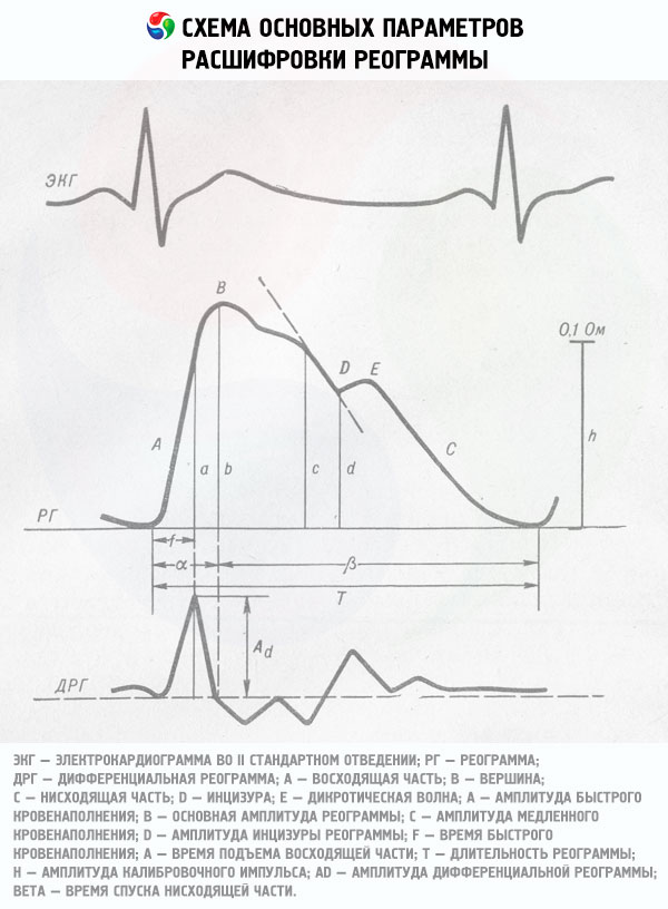
The rheogram resembles a pulsogram in shape. A single REG wave has a beginning, a peak (systolic wave) and an end. The section of the curve from the beginning to the peak is called the ascending (anacrotic) part, the section from the peak to the end of the wave is called the descending (catacrotic) part. Normally, the ascending part is shorter and steeper, and the descending part is longer and flatter. On the descending part, as a rule, one additional wave (dicrotical tooth) is revealed, consisting of a trough and a peak. This complex is called the diastolic wave.
Rheoencephalogram in pathology
Since the configuration of the REG wave components is largely determined by the reflection of the pulse wave from the branching points of the arteries, as well as the elasticity and tone of the vascular wall, changes in the shape of the REG can be used to judge certain disturbances in cerebral blood flow.
With an increase in vascular tone, the amplitude decreases and the top of the systolic wave flattens, the additional (diastolic) wave shifts toward the top, and the severity of the depression decreases. With a decrease in vascular tone, on the contrary, there is an increase in the amplitude and sharpening of the systolic wave, an increase in the severity of the additional wave and its shift toward the end of the REG wave.
When venous outflow is obstructed, the REG curve flattens out and becomes dome-shaped, and with venous hypotension, a small pre-systolic wave appears before the onset of the systolic wave.
The software of modern computer rheographs allows automatic measurement of the listed amplitude-time parameters of the REG wave, as well as calculation of a number of special indices describing the relationships between them, which are more informative for assessing the tone and resistance of large, medium and small arteries and veins than the absolute values of the REG parameters.