Medical expert of the article

New publications
Joint synovectomy
Last reviewed: 06.07.2025

All iLive content is medically reviewed or fact checked to ensure as much factual accuracy as possible.
We have strict sourcing guidelines and only link to reputable media sites, academic research institutions and, whenever possible, medically peer reviewed studies. Note that the numbers in parentheses ([1], [2], etc.) are clickable links to these studies.
If you feel that any of our content is inaccurate, out-of-date, or otherwise questionable, please select it and press Ctrl + Enter.
If conservative treatment of certain joint diseases does not produce results, an operation is performed to remove the damaged part or the entire synovial membrane lining the joint capsule – synovectomy.
Removing abnormal tissue can reduce symptoms and slow the destruction of joint cartilage. [ 1 ]
Indications for the procedure
In orthopedic surgery, synovectomy is used when symptoms of synovial membrane alteration in the joint, such as severe pain and disability-threatening limited mobility, do not respond to either medication or physical therapy for at least 10-12 months. [ 2 ]
And the main indications for the removal of synovial tissue are the presence of radiologically confirmed:
- rheumatoid arthritis; [ 3 ]
- seronegative spondyloarthropathies, including reactive and psoriatic arthritis;
- septic arthritis;
- post-infectious or trauma-induced monoarthritis;
- synovitis (including infectious);
- synovial tumor - pigmented villonodular (villous-nodular) synovitis;
- recurrent hemarthrosis (developing joint damage in patients with hemophilia); [ 4 ]
- chronic form of aseptic bursitis.
Limited and sometimes total synovectomy is used in cases of relapse of primary synovial osteochondromatosis (formation of osteochondral bodies in the synovial membrane).
As for synovectomy in rheumatoid arthritis, as foreign specialists note, this procedure for pain relief in case of damage to the knee or elbow joint (accompanied by synovitis) can be used provided that the destruction of bone or cartilage is minor. But if the cartilage is severely damaged and the destruction of the joint progresses rapidly, synovectomy will not help. In such cases, joint replacement ( endoprosthetics ) is required.
Preparation
In preparation for synovectomy, the surgeon studies the patient's medical history and available images of the affected joint, conducts a physical examination, and also prescribes instrumental studies: X-ray, computed tomography (CT) and MRI scanning - to confirm the existing disease, as well as detailed visualization of all bone and connective tissue structures of the joint and periarticular tissues at the time of surgery.
Standard laboratory tests before surgery include a coagulogram – a blood test to determine how well your blood clots.
Technique synovectomies
The technique used depends on the method by which the synovectomy is performed, and the choice of method is determined by the specificity and degree of damage to the joint and its localization.
Thus, on the upper limbs (most often with arthritis), synovectomy of the wrist, elbow and shoulder joints is performed; on the lower limbs – synovectomy of the ankle, knee and hip joints (especially the acetabulum).
According to clinics, the majority of patients undergo knee synovectomy, with elbow synovectomy coming in second place.
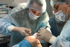
Open surgery (arthrotomy) and arthroscopic are the two main methods of removing synovial tissue, and both are performed under general anesthesia. [ 5 ]
In open synovial ablation surgery, an incision is made over the affected joint, the joint capsule is exposed and dissected, and the inflamed or pathologically altered synovial membrane is scraped or excised, and the effusion is removed. In cases of bone infection, the joint is sanitized. The incisions are sutured, and a bandage is placed on top of the joint.
During arthroscopic synovectomy, several small percutaneous incisions (portals) are made around the joint perimeter using trocars, through which an arthroscope (a flexible tube equipped with a light guide and a video camera) and miniature surgical instruments are inserted. Before removing the synovial membrane, a sterile solution is injected into the joint capsule through a cannula. The surgeon performs all manipulations while looking at the magnified image received from the arthroscope camera on the monitor. At the end of the procedure, all surgical devices are removed, and a bandage is applied to the incisions. [ 6 ]
Experts note such obvious advantages of arthroscopic technique (especially in synovectomy of the shoulder and knee joint) as minimal trauma to periarticular tissues, absence of kinesthesia disorders, less pronounced postoperative pain and faster recovery of patients. [ 7 ]
Although arthroscopy is less invasive than open surgery, the technique is more complex and the procedure takes longer.
Contraindications to the procedure
Synovectomy is not performed:
- for osteoarthritis and ostearthritis;
- in the acute stage of joint inflammation of infectious etiology;
- in the presence of progressive rheumatoid arthritis with radiologically determined high degree of joint destruction (subchondral bone and/or articular cartilage);
- in cases of severe joint instability;
- in case of ankylosis.
Also on the list of contraindications are severe ischemic heart disease, pregnancy and breastfeeding.
Consequences after the procedure
Since standard synovectomy results in regeneration of the synovial membrane of the joint over time (due to the formation of connective tissue during the maturation of fibroblasts), the most common consequence after the procedure is a relapse of synovitis or chondromatosis and even their progression – with the need for a repeat operation. [ 8 ]
According to some data, almost 15-20% of patients who underwent arthroscopic synovectomy of the hip joint experience relapses of synovial chondromatosis within the first two to three years after the procedure.
Complications after the procedure
The main complications after synovectomy are associated with a negative reaction to anesthesia, infection and the development of an inflammatory process, damage to blood vessels and bleeding, damage to nerves, and damage to the surfaces of articulating bones. [ 9 ]
As clinical experience shows, there is a high risk of nerve damage during synovectomy of the elbow joint; during open synovectomy of the shoulder joint, coordination of the muscles of the shoulder and shoulder girdle may be impaired; in some patients after synovectomy of the ankle joint, due to scars and contracture, mobility of the limb at the ankle is significantly reduced.
Moreover, open synovectomy more often than arthroscopic one leads to postoperative joint rigidity and a decrease in its range of motion.
Care after the procedure
Postoperative care and subsequent rehabilitation are carried out according to the instructions and recommendations of the surgeon who performed the operation. In particular, regarding restrictions on joint movement (rotations, extension-flexion, etc.) and the optimal position of the limb: the elbow joint is kept in a bent position (using an orthosis), after surgery on the knee joint, its immobilization is ensured by a removable plaster cast, and the leg should be kept slightly bent (for which a bolster or small pillow is placed under the knee). [ 10 ]
In case of joint swelling, cold is applied; in case of pain, painkillers are prescribed, heparin is used to prevent blood clots, and non-steroidal anti-inflammatory drugs (NSAIDs) are used to prevent ossification.
Postoperative rehabilitation consists of performing a set of exercises determined in each specific case by a specialist (rehabilitation specialist or physiotherapist) taking into account the balance of active and passive movement - to develop joint mobility and restore its functions. And physiotherapy can begin two days after surgery and should continue for at least two or even three months. [ 11 ]
Although the total rehabilitation time depends on the patient's condition and the degree of joint damage. Thus, pain after synovectomy goes away, on average, in three to three and a half weeks; swelling subsides and joint mobility improves noticeably in a month or a month and a half.