Medical expert of the article

New publications
Hypomelanosis
Last reviewed: 05.07.2025

All iLive content is medically reviewed or fact checked to ensure as much factual accuracy as possible.
We have strict sourcing guidelines and only link to reputable media sites, academic research institutions and, whenever possible, medically peer reviewed studies. Note that the numbers in parentheses ([1], [2], etc.) are clickable links to these studies.
If you feel that any of our content is inaccurate, out-of-date, or otherwise questionable, please select it and press Ctrl + Enter.
Hypomelanosis is a pathology of the formation of skin pigmentation against the background of some disease. The development of hypomelanosis is based on a violation of the production of melanin by melanocytes located in the thickness of the skin. This pathological condition can manifest itself in the form of leukoderma, a reduced amount of melanin, as well as its complete absence.
The trigger for the development of hypomelanosis is damage to one or more links in the production and conversion of melanin. This may be the absence of melanocytes in the skin, a violation of the formation of full-fledged melanosomes and their transportation to keratinocytes.
The main clinical manifestation of the pathology is considered to be white spots that appear as a result of a previous illness with superficial dyschromia of the skin. Hypomelanosis is often observed among children, which occurs soon after the illness.
To establish a correct diagnosis, a histological examination is often used. After all, without it, hypomelanosis can be missed, which leads to developmental delays in childhood. Therapeutic goals for pathology are aimed at peeling procedures and the use of retinoids.
 [
[ Causes of hypomelanosis
The appearance of white spots can occur in the first days of a baby's life, because the causes of hypomelanosis are genetic. Thus, there is a failure in the synthesis of melanin - a special pigment responsible for the color of the skin.
The production of melanin begins due to the action of a special enzyme - tyrosinase, after which many chain reactions are launched at the molecular level. This complex process is regulated by a special combination of genes, among which the breakdown occurs.
Thus, the causes of hypomelanosis should be sought in the genetic apparatus. In addition, the transmission of pathology is characterized by a recessive type, especially among consanguineous marriages. The carrier of the gene can be exposed by the presence of a patch of gray hair with clear boundaries, freckles and white spots on the skin.
Due to the fact that the exact causes of hypomelanosis are not clear, and genetic breakdowns cannot be influenced yet, therefore there is no pathogenetic treatment. Thanks to research, it was possible to discover methods and drugs that can partially normalize melanin synthesis.
Symptoms of hypomelanosis
Due to the fact that this pathological condition has genetic causes of impaired melanin production, the first clinical manifestations of hypomelanosis can be observed from the birth of the baby.
Symptoms of hypomelanosis are expressed by the formation of a white area of skin with clear boundaries, which differs from the shade of the rest of the skin. The number and size of such spots can vary and increase over time.
If the baby has pale or white skin, then the symptoms of hypomelanosis may not be immediately noticeable. For more accurate visualization, a Wood's lamp is required to examine the area without pigmentation in a dark room.
This lamp enhances the contrast between the normal color of the skin and hypomelanosis. In the case of the development of Ito's hypomelanosis, in addition to skin manifestations, the development of a pathology of the nervous system with neurological disorders in the form of mental disorders and increased convulsive readiness is possible, and anomalies of the skeletal system are also observed.
Hypomelanosis in a child
Insufficient pigment production in babies may indicate the presence of Wardeburg syndrome, which is transmitted genetically in a dominant manner. Its main clinical manifestations are considered to be white strands of hair, areas of hypopigmentation on the skin, different colors of the iris and eye levels, as well as a wide bridge of the nose and congenital deafness.
In addition, hypomelanosis in a child is observed with tumorous sclerosis, which is characterized by the appearance of white spots up to 3 cm in size and localized on the body, as well as nodules on the forehead, arms and legs. In addition to skin manifestations, mental retardation, epilepsy, phacomatosis of the retina, cyst-like formations in the kidneys, lungs, bones and cardiac rhabdomyomas are observed.
Hypomelanosis in a child is observed with hypomelanosis Ito with the appearance of hypopigmented areas of the skin of various shapes in the form of waves and stripes. Such symptoms may disappear on their own with age.
Vitiligo is also a defect of pigment synthesis, which is characterized by the appearance of white skin areas with a clear outline. Localization is possible on the face, genitals, feet, hands, in the area of joints.
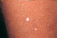
Guttate hypomelanosis
This form of pathology can most often be observed in female representatives of the population aged 35-55 years. Women with a light skin tone and those who spend a long time in direct sunlight are most susceptible to hypomelanosis.
As a result, the number of melanocytes in the affected areas decreases by almost 2 times. In addition, there are opinions that guttate hypomelanosis is associated with HLA-DR8.
Genetic factors play an important role in the development of this disease, especially if it is observed in close relatives.
Clinical manifestations of hypomelanosis are characterized by the appearance of white, round spots on the skin. The diameter of such altered areas reaches up to 1 cm.
Guttate hypomelanosis first appears on the shin (extensor surface), and then spreads to the forearms, upper back and chest. This pathological condition does not typically involve the skin of the face.
The progression of the process is observed with age, as well as with excessive exposure to direct sunlight.
Hypomelanosis of Ito
The pathology is observed in men and especially women and is second only to neurofibromatosis and tuberous sclerosis in prevalence. Hypomelanosis Ito is a sporadic disease, but recessive and dominant inheritance are not excluded.
The development of the pathology is based on a failure of cell migration from the neural tube during the intrauterine period, which results in an abnormal arrangement of gray matter in the brain, as well as an insufficient number of melanocytes in the thickness of the skin.
Migration of melanoblastomas occurs in the second and third trimesters of pregnancy. At the same time, neuronal migration is observed, resulting in hypomelanosis ito including clinical manifestations of pigmentation disorders and brain pathology.
Skin symptoms are expressed by areas of hypopigmentation of irregular shape (curls, zigzags, waves). Most often, these lesions are localized near the Blaschko lines, and their appearance can be observed already in the first days or months of the baby's life, but by adolescence they may become less noticeable or disappear altogether.
Neurological symptoms are characterized by mental disorders, epileptic seizures, which are distinguished by their resistance to anticonvulsant therapy. Often, children suffer from autism, muscle hypotonia and motor disinhibition. Macrocephaly is noted in a quarter of cases.
In addition, pathology of other organs is often observed, for example, heart defects, abnormalities in the structure of the genitals, face, deformations of the spine, feet, eye symptoms, as well as abnormalities in the structure and growth of teeth and hair.
Idiopathic hypomelanosis
The development of hypomelanosis is based on a disruption in the stages of melanin synthesis due to the absence of melanocytes, a failure in the formation of full-fledged melanosomes and their migration.
Melanocytes originate from ectomesenchyme. Their differentiation goes through 4 stages. The first is the appearance of melanocyte precursors in the neural crest, the second is the migration of melanocytes in the thickness of the dermis towards the basement membrane of the epidermis. Then their movement in the epidermis itself is noted and, finally, the stage of formation of processes (dendritic), when the cell takes its position in the epidermis.
Idiopathic hypomelanosis develops in the event of a breakdown at one of the listed stages, as a result of which the melanocyte can be located in an unusual place for it, due to which a certain area of the skin remains “colorless”, since pigment synthesis will be absent.
It can manifest itself in babies or with age. In addition, when exposed to ultraviolet rays, this pathology may progress.
It is quite difficult to identify the main cause of the disease, since in almost 100% of cases it is a genetic defect. Idiopathic guttate hypomelanosis can manifest itself immediately after birth or in adolescence. Most often, the pathology has a chronic type of course with periodic relapses.
Clinical manifestations of the disease are foci of hypopigmentation of various localizations (shins, forearms, back) and up to 1 cm in diameter. The areas are located separately from each other and are not capable of merging.
Most often, idiopathic guttate hypomelanosis is observed in women with a light skin tone, especially those living in areas with increased exposure to sunlight. In addition, when the lesion first appears on the shin, then under the influence of insolation, the number of depigmented areas increases.
There is no pathogenetic therapy aimed at removing the causative factor, therefore symptomatic treatment is used to reduce the intensity of the manifestations of the pathology.
Diagnosis of hypomelanosis
Violation of pigmentation processes can manifest itself in various forms. To verify the pathology, in addition to visual inspection, it is necessary to use a Wood's lamp study. It is especially often used in the presence of light skin and unclearly manifested pathology.
The diagnosis of hypomelanosis is based on the identification of clear boundaries of the hypopigmented lesion by shining a lamp in a darkened room. Thanks to it, it becomes possible to detect the area and verify it.
Diagnosis of Ito hypomelanosis additionally includes a computed tomography scan of the brain, which reveals an enlargement of the 3rd and lateral ventricles, blurred boundaries between brain matter, and atrophy of the frontal lobes.
Histological examination reveals an insufficient number of melanocytes in the hypopigmented area. In addition, with Ito's hypomelanosis, other features may be present in the lesion, such as vascular nevi, cocoa spots, nevus of Ott, or Mongolian blue spots.
How to examine?
Who to contact?
Treatment of hypomelanosis
This pathological process is characterized by its spread at the genetic level, and therefore pathogenetic treatment does not yet exist. Symptomatic therapy is used, the main directions of which are to stop the generalization of the pathology and reduce its clinical manifestations.
Treatment of hypomelanosis involves the use of corticosteroids, which are administered intralesionally. In addition, various studies confirm the effectiveness of topical retinoids, pimecrolimus (Elidel), and cryomassage of the affected areas.
Hypomelanosis can also be treated with phototherapy, which activates melanin production by pigment cells. In addition, replacement therapy with the drug melagynin is recommended. Its action is aimed at stimulating melanocytes to synthesize pigment.
As for bioresonance therapy, it is aimed at restoring the normal functioning of the nervous system, as well as increasing the level of the body's immune forces.
Traditional treatment is also possible for this type of pathology, but before using it, be sure to consult a doctor.
Prevention of hypomelanosis
There is no specific prevention of hypomelanosis, since the pathology has a genetic type of inheritance. However, there are still methods that can reduce the risk of developing hypomelanosis or its recurrence.
The main provoking factor of the generalization and progression of the process is considered to be excessive insolation. As a result, it is necessary to inform the population about its negative impact not only in relation to hypomelanosis, but also the likelihood of developing cancer.
Prevention of hypomelanosis consists of avoiding exposure of unprotected skin to direct sunlight, especially between 11:00 and 16:00, using sunscreen cosmetics in hot weather, as UV radiation can be reflected from surrounding objects and the ground, pass through clouds and clothing. As a result, it is not recommended to be outdoors during the daytime unless absolutely necessary. This also applies to those who like to tan in a solarium. To protect the skin, it is necessary to use special creams, a hat and clothing that covers areas of hypomelanosis.
Prognosis of hypomelanosis
The hypopigmentation areas in the form of white spots themselves do not pose a health threat, but when the first clinical symptoms appear, you should consult a specialist for further diagnosis and treatment. The sooner the pathology is detected, the greater the likelihood of stopping the process and preventing the development of relapses.
The prognosis for hypomelanosis is favorable, but with excessive exposure to sunlight, it can spread to healthy tissues, since excessive insolation contributes to a decrease in the number of melanosomes and pigment.
It is impossible not to warn about the possibility of developing a carcinogenic process under the influence of sunlight. This is due to the malignant degeneration of cells due to disturbances in gene expression. In addition, each person has birthmarks, which are also capable of changing under the influence of the sun.
So, hypomelanosis is not a terrible pathology, but still requires a special approach and adherence to specific measures to prevent occurrence and relapse during a chronic course.