Medical expert of the article

New publications
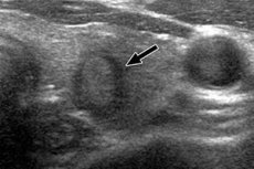
Hypoechogenic mass
Last reviewed: 04.07.2025

All iLive content is medically reviewed or fact checked to ensure as much factual accuracy as possible.
We have strict sourcing guidelines and only link to reputable media sites, academic research institutions and, whenever possible, medically peer reviewed studies. Note that the numbers in parentheses ([1], [2], etc.) are clickable links to these studies.
If you feel that any of our content is inaccurate, out-of-date, or otherwise questionable, please select it and press Ctrl + Enter.
Instrumental diagnostics using ultrasound scanning (ultrasound), which is also called ultrasonography, can reveal areas with different acoustic density in internal organs and cavities – hyperechoic or hypoechoic formations.
What does hypoechoic lesion mean?
A local hypoechoic formation in a particular organ, unlike a hyperechoic one, is the result of lower tissue echogenicity – in comparison with the acoustic density parameters of healthy tissues of the organ. That is, this area weakly reflects the ultrasound signal directed at it (in the frequency ranges of 2-5, 5-10 or 10-15 MHz). And this is evidence that this formation – from the point of view of its structure – either contains liquid or has a cavity.
Hypoechoic formations are visualized on the screen as gray, dark gray, and almost black zones (with hyperechogenicity, the zones are light, often white). To decipher the ultrasound image, there is a scale of six gray categories Gray Scale Imaging, where each pixel of the image of the hypoechoic formation obtained on the monitor - depending on the strength of the ultrasound signal returning to the sensors - represents a specific shade of gray.
The results of the ultrasound examination, deciphered by ultrasound diagnosticians (sonographers), are studied by doctors of a specific profile (endocrinologist, gastroenterologist, urologist, nephrologist, oncologist, etc.), compared with the results of tests taken by patients and the results of other studies.
In many cases, differential diagnostics is required, for which, in addition to ultrasound, other hardware methods of visualizing pathology are used (angiography, color Doppler, CT, MRI, etc.), and histological examination of biopsies is also carried out.
Causes of hypoechoic formation
As an ultrasonography indicator, a hypoechoic formation can have any localization. The causes of a hypoechoic formation are also different and completely depend on the etiology and pathogenesis of the diseases that develop in patients.
For example, a hypoechoic formation in the pancreas is considered a diagnostic criterion for identifying pathologies such as cysts, hemorrhagic pancreatitis, mucinous cystadenoma (which is prone to malignancy), adenocarcinoma of the head of the pancreas, and metastases from malignant tumors of other organs.
Hypoechoic formation in the liver and gallbladder
Healthy liver tissues are moderately hyperechoic, and a hypoechoic formation in the liver may occur with cirrhotic foci; focal steatosis; cysts (including Echinococcus multilocularis); biliary abscess; hepatocellular adenoma; focal parenchymal hyperplasia; hepatoma and small cholangiocellular adenocarcinoma.
Hypoechoic formations are also visualized in cases of spread of diffuse metastases to the liver from pancreatic, ovarian, mammary gland, testicular, and gastrointestinal cancers.
In ultrasound diagnostics of gallbladder pathologies, the structure of its walls is of particular importance, since in the absence of organ damage they are visualized as three layers: external and internal hyperechoic and middle hypoechoic.
Among the causes of hypoechoic formation in the gallbladder are polyps, adenocarcinoma (with an intact outer layer of the bladder), lymphomas (tumors of the lymph nodes), and angiosarcoma.
Hypoechoic lesions of the spleen
Normally, the echogenicity of the spleen is uniform, although slightly higher than that of the liver. However, due to high vascularization, ultrasound of the spleen is performed with a contrast agent that accumulates in the parenchyma and makes it possible (at the end of the parenchymatous phase) to visualize focal lesions and hypoechoic formations of the spleen.
These formations include:
- acute intraparenchymal hematoma due to rupture of the spleen (due to abdominal trauma);
- hemangiomas (benign vascular formations) with splenomegaly;
- splenic infarctions (infiltrative or hematological);
- splenic lymphoma;
- metastases of various origins (most often soft tissue sarcomas, osteosarcomas, kidney cancer, breast cancer or ovarian cancer).
As experts note, echinococcal, tapeworm and dermoid cystic formations of the spleen can have a mixed echostructure.
Hypoechoic formation in the kidney, adrenal glands and bladder
A hypoechoic formation in the kidney can be detected when cystic formations (including malignant ones), hematomas (in the initial stages), pyogenic paranephric abscesses (at the necrosis stage) or cavernous tuberculosis of the kidney are included in the parenchyma.
According to endocrinologists, detecting a hypoechoic formation in the adrenal gland is not an easy task, and ultrasound, unfortunately, does not always cope with it. For example, verification of the diagnosis of adenoma in primary aldosteronism, as well as pathological proliferation of adrenal cortex cells in hypercorticism (Itsenko-Cushing's disease) is based on symptoms. Ultrasound accurately detects a fairly large pheochromocytoma, as well as lymphoma, carcinoma and metastases. So, it is most advisable to examine the adrenal glands using CT and MRI.
In the development of benign leiomyoma, transitional cell carcinoma of the bladder or pheochromocytoma (paraganglioma) of the bladder, which is accompanied by arterial hypertension and hematuria, a hypoechoic formation in the bladder is visualized during an ultrasound examination.
Hypoechoic formation in the abdominal cavity and pelvis
Pathologies localized in the abdominal cavity, in particular in the intestinal section of the gastrointestinal tract, are easily examined by ultrasound: a diseased empty intestine has thickened hypoechoic walls that contrast with the surrounding hyperechoic adipose tissue.
The far from complete list of reasons causing a hypoechoic formation in the abdominal cavity visualized by ultrasound includes:
- a hernia protruding into the inguinal canal;
- intra-abdominal hematomas (traumatic or associated with coagulopathy);
- serous and purulent phlegmon of the peritoneum or retroperitoneal space;
- abscess of the terminal ileum in transmural ileitis (Crohn's disease);
- inflammation of the mesenteric lymph nodes (lymph nodes of the mesentery);
- B-cell non-Hodgkin lymphoma or Burkitt lymphoma;
- metastasis to the visceral lymph nodes of the abdominal cavity;
- carcinoma of the cecum, etc.
During ultrasound examination of the pelvic organs and uterus, formations with low acoustic density are detected in women - in the presence of myoma, adenoma, cyst or endometriosis of the uterus; functional or dermoid cysts of the appendages. And a hypoechoic formation in the ovary occurs with a hemorrhagic cyst, as well as tubo-ovarian abscess (purulent inflammation in the fallopian tubes and ovaries), follicular lymphoma and carcinoma.
In men, pathologies with such a diagnostic indicator are testicular cancer, testicular lymphocele, varicocele of the cord, and during an ultrasound examination of the prostate in patients with benign adenoma or cancer of this gland, a hypoechoic formation of the prostate gland is visualized.
Hypoechoic formation in the subclavian region
A hypoechoic formation in the subclavian region detected during an ultrasound scan may be a sign of:
- benign neoplasms and malignant lymphomas of the anterior mediastinum;
- chronic lymphocytic leukemia;
- lesions of peripheral lymph nodes by metastases of thyroid cancer, larynx, esophagus, mammary glands, lungs;
- osteosarcoma of thoracic localization;
- cysts and echinococcosis of the lungs;
- thymomas or carcinomas of the thymus gland.
Hypoechogenicity of structures in this area is noted by clinicians in patients with hyperplasia or cysts of the parathyroid glands, hyperparathyroidism or nodular adenomatosis.
Types of hypoechoic formations
In addition to the anatomical and topographic characteristics of the resulting formation, ultrasonography reveals its shape (round, oval, irregular), size in width (craniocaudal) and depth relative to the outer wall of the organ or cavity.
According to this parameter, the main types of hypoechoic formation include:
- a round hypoechoic formation or a hypoechoic oval formation (these are various cysts, varicoceles, adenomas, adrenal tumors of metastatic etiology);
- hypoechoic nodular formation (characteristic of hemangiomas, nodular biliary hypertrophy, uterine fibroids, nodular adenomatosis, etc.);
- hypoechoic focal formation (characteristic of cirrhosis and focal fatty infiltration of the liver, hematomas and infarction of the spleen, etc.).
The conclusion of the ultrasound examination notes the features of the image contours:
- hypoechoic formation with smooth contours (cysts, nodular hypertrophy of the liver, breast tumors );
- hypoechoic formation with uneven contours (many tumors, most metastases);
- hypoechoic formation with a clear outline (cysts, adenomas, abscesses that have a hyperechoic rim on the ultrasound image);
- hypoechoic formation with unclear contours (cavernous hemangiomas of the liver, thyroid cancer, metastases in organ tissues of any localization).
Next, the homogeneity/heterogeneity of the formation, that is, its internal structure, is assessed:
- hypoechoic homogeneous formation (carcinoma);
- hypoechoic heterogeneous formation (large adenomas, liver cancer, diffuse forms of carcinomas, etc.);
- hypoechoic formation with hyperechoic inclusions (renal cell carcinoma, ovarian adenoma, prostate cancer).
A description of the state of the surrounding tissues, distal acoustic effects (enhancement, attenuation, acoustic shadow) and the characteristics of lateral shadows (symmetry, asymmetry, absence) is mandatory.
In addition, the presence/absence of vascularization (i.e. blood vessels) in nodular formations is noted, with the definition of such types as: hypoechoic formation without blood flow (avascular) and hypoechoic formation with blood flow.
Formations containing blood vessels are divided into:
- hypoechoic formation with perinodular blood flow (subtype with perinodular, i.e., vascularization surrounding the node);
- hypoechoic formation with combined blood flow (vessels are present near the formation and inside it);
- hypoechoic formation with intranodular blood flow (the presence of vascularization is recorded only inside the formation).
As clinical practice shows, a hypoechoic formation with intranodular blood flow may indicate its malignant nature.
And finally, the presence of calcium compounds in the structure of the formation is taken into account. And a hypoechoic formation with calcifications (calcinosis) is characteristic of encapsulated chronic liver abscess in amebiasis, liver cancer, neoplasms in the thyroid and prostate glands, malignant tumors of the mammary gland, etc.
Who to contact?
Treatment
Patients may ask the doctor what treatment is needed for a hypoechoic formation and what medications are prescribed for it... But they do not treat abnormal areas detected by ultrasound, but diseases that have caused pathological changes in the density of tissue that weakly reflects ultrasound waves.