Medical expert of the article
New publications
Hepatitis G virus (GB-C)
Last reviewed: 08.07.2025

All iLive content is medically reviewed or fact checked to ensure as much factual accuracy as possible.
We have strict sourcing guidelines and only link to reputable media sites, academic research institutions and, whenever possible, medically peer reviewed studies. Note that the numbers in parentheses ([1], [2], etc.) are clickable links to these studies.
If you feel that any of our content is inaccurate, out-of-date, or otherwise questionable, please select it and press Ctrl + Enter.
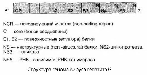
Hepatitis G virus (HGV) was discovered in 1995 and belongs to the Flaviviridae family (Hepaciviridae genus). The G virus genome is a single-stranded, non-fragmented, positive-sense RNA 9,500 bases long. The structural organization of the G virus genome is similar to that of HVC. The genome contains one large reading frame that encodes a precursor polyprotein containing about 2,800 amino acid residues. It is cut by cellular and viral proteases to form two structural and at least five nonstructural proteins. The genes encoding the structural proteins (cor and env) are adjacent to the 5' end of the viral RNA, and the genes encoding the nonstructural proteins (helicase, protease, polymerase) are adjacent to the 3' end. It has been established that the nonstructural genes of HGV are similar to the genes of the hepatitis C virus, as well as the GBV-A and GBV-B viruses. All these viruses are classified into one genus Hepacivirus of the Flaviviridae family.
In terms of the structure of the structural genes, HGV has nothing in common with GBV-A and HCV and only vaguely resembles GBV-B. The hepatitis G virus turned out to be identical to the GBV-C virus, which was also isolated during the study of a subpopulation of GBV viruses from tamarin monkeys, through which the RNA virus from a patient with acute hepatitis of unknown etiology, with the initials GB, was passaged; in his honor, all these viruses were named hepatitis viruses GBV-A, GBV-B, GBV-C. The HGV virus (GB-C) has a defective cor protein and exhibits less pronounced variability than HCV. Three types and five subtypes of the HGV genome have been identified. Genotype 2a dominates, including in Russia, Kazakhstan and Kyrgyzstan.
HGV RNA is constructed according to a pattern characteristic of the entire flavivirus family: at the 5' end is a zone encoding structural proteins, and at the 3' end is a zone encoding non-structural proteins.

The RNA molecule contains one open reading frame (ORF); it codes for the synthesis of a precursor polyprotein consisting of approximately 2900 amino acids. The virus has constant regions of the genome (used to create primers used in PCR), but is also characterized by significant variability, which is explained by the low reliability of the reading function of the viral RNA polymerase. It is believed that the virus contains a core protein (nucleocapsid protein) and surface proteins (supercapsid proteins). Different variants of capsid proteins have been detected in different isolates; it can also be assumed that defective capsid proteins exist. Different variants of the nucleotide sequences of HGV in different isolates are regarded as different subtypes within a single genotype or as intermediate between genotypes and subtypes. At the same time, some authors believe that there are different genotypes of HGV, including GBV-C and HGV-prototype among the latter.
Markers of the G virus are found in 2% of the population of these countries. The G virus is found in 1-2% of blood donors in different countries of the world, i.e. more often than the hepatitis C virus. Like the hepatocyte viruses HBV/HCV, this virus is capable of persistence, but less often leads to chronic pathology, and this persistence probably proceeds as a healthy carrier. Acute clinical manifestations of hepatitis G are also less severe than those of hepatitis B and C. CPR and IFM are used to diagnose hepatitis G.


 [
[