Medical expert of the article

New publications
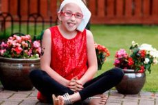
Goldenhar syndrome
Last reviewed: 04.07.2025

All iLive content is medically reviewed or fact checked to ensure as much factual accuracy as possible.
We have strict sourcing guidelines and only link to reputable media sites, academic research institutions and, whenever possible, medically peer reviewed studies. Note that the numbers in parentheses ([1], [2], etc.) are clickable links to these studies.
If you feel that any of our content is inaccurate, out-of-date, or otherwise questionable, please select it and press Ctrl + Enter.
Oculo-auriculo-vertebral dysplasia, as this rather rare congenital pathology is also called, usually affects the development of organs of one half of the face: eyes, ears, nose, soft palate, lips, jaw. It is a separate type of facial microsomia, in which, due to intrauterine underdevelopment of skeletal, neuromuscular and other components of soft tissues (derivatives of the first and second gill slits), the external organs of one half of the face are noticeably smaller in size. Very rarely, this pathology is bilateral.
Epidemiology
Medical statistics show that diseases of the oculo-auriculo-vertebral spectrum in the structure of intrauterine anomalies of the craniofacial zone follow such pathologies as cleft lip and cleft palate, both combined and separate. The frequency of Goldenhar syndrome, mentioned in foreign medical literature, is one child out of 3.5-7 thousand live births. This syndrome is diagnosed in one out of a thousand deaf newborns. In approximately 70% of cases, the lesions are unilateral, with bilateral defects they are more pronounced on one side, and among them the right side prevails with an incidence rate of 3:2. Distribution by gender - for every three male children there are two girls.
Causes of Goldenhar syndrome
The combination of dysplasia of the eyes, ears and spine, described in the early 1950s by the American doctor M. Goldenhar, was immortalized by his name. This rare congenital pathology has not yet been studied much, but the opinion of most researchers coincides in that it is etiologically caused by a genetic predisposition. The type of inheritance is currently not determined, cases of the disease are sporadic. There are reports of autosomal dominant familial inheritance. Patients with this disease have chromosomal abnormalities. Hypothetically, the risk factors for the birth of a child with dysplastic lesions of the oculo-auriculo-vertebral structures are consanguineous marriage, and abortions in the mother before the birth of the child, and teratogenesis of an exogenous or endogenous nature, in particular, diabetes mellitus or alimentary obesity of the expectant mother.
The risk of having a sick child in a genetic carrier is 3%, and the recurrence of having a child with this defect in one family is equal to 1%.
 [ 9 ]
[ 9 ]
Pathogenesis
Pathogenesis, again hypothetically, is based on the possibility of hemorrhage in the area of the first and second gill slits of the embryo, coinciding in time with the replacement of the source of blood supply in this area. The blood supply from the stapedial artery is replaced by the supply from the external carotid artery. Vascular stroke occurring at this time and in this place leads to pathological transformations of cell proliferation and abnormal formation of bone-muscle, nerve and other elements of soft tissues developing from the derivatives of the first and second gill slits.
Symptoms of Goldenhar syndrome
The first signs of facial microsomia are mainly determined visually during examination of the newborn. Typical symptoms are some facial asymmetry, abnormal size and position of the eye socket, deformation of the auricles in the form of specific auricular "protrusions", while other changes in the outer ear may be absent, underdevelopment of the lower jaw.
As the child grows, the symptoms become more noticeable. The Goldenhar phenotype includes developmental abnormalities of the ear (microtia), eyes, nose, soft palate, lips and jaw. One of the typical symptoms is the presence of choristomas (epibular dermoids) on the surface of the eyeball. These are tumor formations containing tissues that are not typical for their localization (hair follicles, sebaceous and sweat glands, fibrofatty tissue). This symptom is specific for 70% of Goldenhar syndrome cases. Ophthalmologic malformations may include (25% of cases or more) lipodermoids in the outer conjunctiva of the eyeball, columbae of the upper eyelid, defects of the oculomotor muscles, and an eye shape with the outer corners of the eyeballs drooping downwards. Rarely (no more than 5% of cases) a small diameter of the cornea of the eye, a through defect or absence of the iris, drooping of the upper eyelid, underdevelopment of the eyeball and its small size, strabismus and cataracts are observed.
Developmental anomalies of the auricles are the most common. They are deformed and noticeably smaller than normal in size (approximately 80% of patients), half of the patients with the syndrome have an abnormal location, and the external auditory canal may be absent (40% of patients). 55% of patients had defects in the development of the middle ear and hearing loss.
A very characteristic sign of Goldenarch syndrome (85%) is underdevelopment of the lower jaw processes, and the facial muscles, upper and lower jaws are also quite often asymmetrical and underdeveloped. When examining the oral cavity, a high-arched palate is observed, sometimes with a cleft, open bite, too wide oral slit, cleft tongue and additional frenulum.
Slightly less than half of the cases were accompanied by underdevelopment of the vertebrae, most often in the cervical region - wedge-shaped, fused, hemivertebrae, scoliosis, a third - spina bifida, rib malformations, a fifth - clubfoot.
Less than a third of Goldenhar syndrome cases were accompanied by cardiovascular abnormalities (ventricular septal defect, patent ductus arteriosus, tetralogy of Fallot, narrowing or complete occlusion of the aorta). Mental retardation of varying degrees was observed in a tenth of patients with this syndrome.
There are several classifications that reflect the stages of the disease, or rather the degree of its severity. The most complete is OMENS. It identifies three stages of severity of damage to each of the objects of malformations in hemifacial microsomia: eyes (orbit), lower jaw (mandible), ear (ear), facial nerve (facial nerve) and bones of the skeleton (skeletal). Since the defects are multiple and each structure is usually affected to varying degrees, it looks something like this: O2M3E3N2S1*. The asterisk reflects the presence of additional defects of non-craniofacial objects.
The SAT classification focuses on three main objects: the skeleton, the auricle, and soft tissue. According to this classification, skeletal malformations are considered in five stages (from S1 to S5), auricular structure disorders – in four (from AO to A3); soft tissue defects – in three (from T1 to T3). Thus, the mildest stage of the disease is S1A0T1, severe malformations – S5A3T3. The SAT system is inferior to the previous one in the absence of important lesions, which are not reflected in it.
Some authors distinguish types of hemifacial microsomia by the phenotype associated with the objects of damage. In this classification, the Goldenhar type is distinguished as a separate type with its specific developmental defects.
Complications and consequences
The consequences and complications of this congenital pathology are directly dependent on the severity of the malformations, some of which are incompatible with life, while others, such as malocclusion, can cause a number of inconveniences. Much depends on timely treatment. If time is lost, then a child with this pathology will develop hypoplasia of the facial bones, which is progressive and increasingly noticeable. It becomes difficult to swallow and chew. Vision and hearing pathologies will also progress. The result of all the deteriorations will be serious physical inconveniences and psychological discomfort, which will affect the quality of life of the child and his parents.
Diagnostics of Goldenhar syndrome
As a rule, a preliminary diagnosis of this congenital anomaly is established in a newborn when facial asymmetry is detected in combination with other specific visual symptoms.
To clarify the diagnosis of this disease, various diagnostic procedures are used. One of the first is to determine hearing acuity, since lesions of the outer and inner ear occur in almost every case and are the first to attract attention. Early hearing examination is caused by the need to prevent the child from lagging behind in psychoverbal development. At an early age, the child is diagnosed during sleep. The following methods are used: impedancemetry, registration of auditory evoked potentials (electrocochleography, otoacoustic emission), computer audiometry.
Older children are tested in a playful manner using speech audiometry. Instrumental and subjective hearing diagnostics are recommended to be performed every six months for seven years.
The primary consultations should be with a maxillofacial surgeon, ophthalmologist, orthodontist, orthopedist, otolaryngologist to diagnose the maximum possible developmental defects. Instrumental diagnostics and tests are prescribed by specialists as needed, depending on the pathologies identified. Electrocardiography, X-ray, and ultrasound examination of internal organs are usually prescribed.
After reaching the age of three, the child is prescribed a computed tomography scan of the temporal zones.
Children with this diagnosis require consultations with many specialists depending on the presence of developmental defects: an audiologist, a speech therapist-defectologist, a cardiologist, a nephrologist, a neurologist, and others.
What tests are needed?
Differential diagnosis
Differential diagnostics are carried out with other congenital craniofacial malformations, such as: dysostoses - mandibular-facial, hemifacial, acrofacial, other types of hemifacial microsomia, Kaufman and orofacial-digital syndromes, Charge's association.
Who to contact?
Treatment of Goldenhar syndrome
The variety of malformations of the skull and spine, as well as other organs and systems in patients with this congenital pathology, leads to multi-stage treatment by many specialists. In mild cases, the child is observed for up to three years, and then surgical treatment begins.
In cases of severe congenital defects, surgical treatment is used first (in infancy or before reaching two years of age). After that, symptomatic complex treatment is carried out. Goldenhar syndrome cannot be cured without multi-stage surgical intervention. The number and scope of surgical operations depend on the severity of the pathologies. Such patients usually undergo compression-distraction osteosynthesis; endoprosthetics of the temporomandibular joint, lower and upper jaw; osteotomy of the nose, lower and upper jaws, correcting their developmental defects and pathological bite; plastic surgery (genioplasty, rhinoplasty). Antibiotic therapy and vitamin therapy are prescribed to prevent inflammatory complications and accelerate the rehabilitation process. Osteotropic antibiotics are usually prescribed for maxillofacial surgical manipulations: penicillins, lincomycin, erythromycin.
Penicillins are natural compounds synthesized by different forms of penicillin mold fungi and semi-synthetic, based on 6-aminopenicillanic acid isolated from natural compounds. Their antibacterial ability is based on the disruption of the bacillus cell membrane. They are low-toxic, have a wide range of dosages, however, most drug allergies are caused by penicillin antibiotics.
Lincomycin is an antibiotic of choice for penicillin allergies, can be prescribed to children from one month of age, therapeutic doses of the drug have a bacteriostatic effect, higher doses have a bactericidal effect, used to eliminate infections of bones, joints, and soft tissues. Contraindicated in severe renal and hepatic dysfunction. May cause allergies.
Erythromycin - belongs to the group of macrolide antibacterial agents, has a wide spectrum of bactericidal action, can be used in ophthalmology. It is prescribed to children from one year. One of the side effects of this drug, as well as a reaction to an overdose, is hearing loss, however, it is considered reversible. Therefore, the drug can be prescribed if the two previous ones are intolerant, especially since prophylactic antibiotic therapy is prescribed for a short time. Its goal is to achieve the highest therapeutic density of the drug in the tissues by the time of surgery. Prophylactic treatment begins an hour or two before surgery and is stopped after two or three days.
Depending on the presence of pain, analgesics are prescribed. Small patients are prescribed children's Nurofen, which has a fast action, and also provides an antipyretic and anti-inflammatory effect and a fairly long action (up to eight hours). The maximum dosage should not exceed 30 mg per kilogram of the child's weight per day.
During the rehabilitation period, it is necessary to provide the child with adequate nutrition and vitamins. Multivitamin complexes are prescribed, including ascorbic acid, retinol, tocopherol, vitamins of group D and B.
To prevent infection and resolve postoperative edema and infiltrates, physiotherapeutic treatment with ultraviolet radiation, ultrasound and electromagnetic waves, as well as laser and magnetic therapy and their combination, and hyperbaric oxygenation are used.
Orthodontic treatment involves the prevention of asymmetric jaw development, correction of abnormal bite, preoperative preparation of teeth and facial muscles for operations. Treatment at the orthodontist is divided into stages corresponding to three types of bite:
- milk - the most important stage of treatment, since the first; the child and his parents are introduced to the pathology, the likelihood of complications and the rules of oral care, devices for correcting jaw defects, and they get used to the necessary procedures:
- replaceable - at this stage, the main task is to correct the bite, prevent and correct developmental defects of the jaws;
- permanent - at this stage, the initiated activities continue, removable devices are replaced with braces, various fixators, depending on the need.
Retention measures, completing the treatment, are carried out to consolidate the achieved results and end at 18 years of age, when the body is almost fully formed. All this time, until reaching adulthood, the child is under the supervision of doctors, depending on the severity of the disease, medications, vitamins and various procedures are prescribed as needed. The treatment complex usually includes therapeutic exercises and work with a teacher of the deaf and a psychologist.
Alternative medicine
Developmental defects of cranial and vertebral structures in Goldenhar syndrome suggest surgical intervention, however, folk treatment can become an additional good help in the rehabilitation period. I would just like to remind you that the possibility of using a folk method must first be discussed with your doctor.
Therapeutic exercises for bite correction
These exercises should be done at least twice a day, each of them repeated at least six times:
- open your mouth as wide as possible, count to ten in this position and close it sharply;
- starting position: touch the tip of the tongue to the palate, move it, without lifting it from the palate, as far back as possible - now open and close your mouth as wide as possible several times;
- sit down, put your elbows on the table, firmly rest your chin on your palms folded horizontally on top of each other - open and close your mouth several times (your lower jaw should be motionless on your palms).
It is also helpful to exercise your jaws by regularly chewing hard foods thoroughly.
Dermoid cysts, characteristic of Goldenarch syndrome, are treated only surgically.
However, there are also folk methods for getting rid of cysts. It is recommended to cleanse the eyes with decoctions of medicinal herbs: for example, make a decoction of cornflower flowers, plantain leaves and caraway seeds, and put three drops in each eye at least five times a day.
You can wash your eyes with tea leaves or a decoction of chamomile flowers: three tablespoons of flowers per glass of boiling water.
Another completely safe and vitamin-rich recipe: mix 1:1 honey and viburnum berry juice. The first week, take one gram of the mixture on an empty stomach in the morning (a level teaspoon contains eight grams of honey), in the second week, double the dose, in the third, double it again, and in the fourth, the dosage is 10 g of honey. Then take a break and repeat the intake in reverse order, starting with 10 g.
Herbal treatment is also used for hearing loss:
- pour 600 ml of boiling water over two teaspoons of calamus root, leave for 2-3 hours, drink three tablespoons before three meals for a month, can be repeated after two weeks;
- Add ½ cup of anise seeds to the top with rosehip oil, leave in a cool, dark place for three weeks; then strain and bury for a month, you can repeat after two weeks;
- Brew and drink rose petal tea without restrictions, it tones the walls of blood vessels and stimulates microcirculation of blood in the ears.
Homeopathy cannot replace surgical interventions required for multiple malformations inherent in Goldenarch syndrome, however, homeopathic preparations can facilitate rapid recovery after surgery (Arsenicum album, Staphysagria). Individual deviations from the norm, such as hearing loss (Asterias rubens), strabismus (Tanacetum), neoplasms on the head, eyelids (Crocus sativus, Graphites, Thuja), as well as the general condition of the patient can be corrected, especially after consultation with a homeopathic doctor.
Prevention
There are no specific measures to prevent this congenital disease. However, a favorable gynecological history of the mother, a healthy lifestyle of both parents and a responsible attitude towards procreation significantly increases the likelihood of having healthy children.
If there is a suspicion of possible pathology, an ultrasound scan of the embryo's face in three dimensions is performed at 20-24 weeks of pregnancy: frontal, horizontal and sagittal. This method of examination gives 100% efficiency. There are also other methods of prenatal diagnostics (fetoscopy, tests), allowing the family to draw a conclusion about the advisability of prolonging the pregnancy.
[ 28 ]
Forecast
With a comprehensive examination and timely detection of all anomalies (preferably in infancy), a responsible attitude of the child's parents and long-term and comprehensive treatment, the prognosis for this pathology is favorable in most cases. In approximately 75% of cases, comprehensive treatment and rehabilitation of children with congenital defects of the skull and face is effective. In some cases, timely treatment and plastic surgery lead to the absence of external signs of the disease. Children study in comprehensive schools, universities, and work as adults.
To the question: How long do people live with Goldenhar syndrome? The answer is: the prognosis for life and health depends fundamentally on the presence of combined defects of other vital organs and systems.