Medical expert of the article

New publications
Gingival necrosis
Last reviewed: 04.07.2025

All iLive content is medically reviewed or fact checked to ensure as much factual accuracy as possible.
We have strict sourcing guidelines and only link to reputable media sites, academic research institutions and, whenever possible, medically peer reviewed studies. Note that the numbers in parentheses ([1], [2], etc.) are clickable links to these studies.
If you feel that any of our content is inaccurate, out-of-date, or otherwise questionable, please select it and press Ctrl + Enter.
Gum necrosis is a pathology that indicates tissue death. Let's consider the main causes of the disease, symptoms, diagnostic methods, as well as treatment and prognosis for recovery.
Necrosis is a pathological form of cell death that leads to the death of tissues and parts of organs in a living organism. The peculiarity of the disease is that in the early stages the symptoms are blurred and difficult to diagnose. Gum necrosis can occur due to injuries, medical manipulations during dental treatment, as well as due to a malfunction of the body due to chronic or acute infection, intoxication, vitamin deficiency and other pathologies.
Exposure to chemicals, high or low temperatures, blood disorders, harmful microorganisms and a number of other factors increase the risk of developing the disease. Many dental diseases contribute to its development. If you have bleeding gums, bad breath and loose teeth, this may indicate the development of necrosis. For example, a disease such as gingivitis, without proper treatment, develops into a chronic form, which causes ulcerative lesions of the gums and, of course, tissue death.
The danger of necrosis is that it is an irreversible process, i.e. lost cells do not regenerate. But if you seek medical help in a timely manner, you can stop the further spread of the disease. If you do not do this, then progressive necrosis leads to a complete loss of chewing function.
Causes of gum necrosis
The causes of gum necrosis are very diverse. The disease may appear due to mechanical impact, trauma, prolonged exposure to cold or high temperatures, or due to pinched blood vessels. Gum tissue death occurs due to disruption of normal blood flow to the cells of the affected area. Very often, gum damage is accompanied by tooth necrosis.
Dentists distinguish traumatic, ischemic, trophoneurotic and toxic origins of gum necrosis. The ischemic form of pathology occurs as a result of circulatory disorders, and the trophoneurotic form occurs due to disruption of the innervation of gum tissue. Let's consider the main causes of gum necrosis:
- Poor oral hygiene leads to bleeding and swelling of the gums, destruction of the attachment of the tooth to the gum and tissue necrosis.
- Regular tissue trauma due to treatment or prosthetics leads to circulatory disorders and gum death. Mechanical trauma due to malocclusion causes inflammation and, in advanced cases, necrosis.
- Hormonal imbalances, blood diseases and a number of other endocrine pathologies lead to dental diseases, which without proper treatment develop into necrosis.
 [ 6 ], [ 7 ], [ 8 ], [ 9 ], [ 10 ], [ 11 ], [ 12 ], [ 13 ]
Arsenic-induced gum necrosis
Gum necrosis from arsenic is a fairly common problem faced by many patients in dental clinics. Arsenic anhydrite in the form of a paste is still used in dental practice. The substance is used for devitalization of the dental pulp. That is, for the removal of the coronal or root pulp with complete destruction of all structures and functions of this tissue. The depth of destruction depends entirely on the dose of arsenic paste and the duration of its action. Such treatment is used for pulpitis (chronic and acute diffuse) with curved or impassable roots in elderly patients, with fibrous pulpitis, limited mouth opening, or allergic reactions to local anesthetics.
Arsenic is a chemical element of the nitrogen family. 5-50 mg of this substance is considered a toxic dose for the human body. The mechanism of toxic action is directly related to metabolic disorders. Since arsenic is a protoplasmic poison, it interacts with sulfhydryl groups. Oxidation processes lead to local tissue destruction. The main targets of arsenic are skin, kidneys, gastrointestinal tract, bone marrow and lungs.
Long-term exposure to arsenic causes toxic changes in periodontitis of the tooth and gum necrosis. If the arsenic paste is not hermetically sealed with a temporary filling, the substance will leak out and cause gum necrosis, and subsequently the death of bone cells, i.e. osteonecrosis. The danger of the latter pathology is that it has a chronic and progressive course. Thus, the duration of the disease can be from 1 to 10 years, and at first the disease proceeds unnoticed. But later the patient suffers from severe bone pain and neuropathy.
Symptoms of gum necrosis
Symptoms of gum necrosis have several stages of development, each of which is characterized by clinical manifestations. Let's consider the main symptoms of necrotic changes in gum tissue:
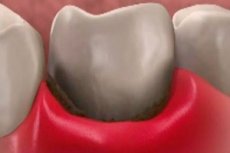
- At an early stage, necrosis may not manifest itself, but it is precisely as the disease develops that the tooth enamel loses pigmentation and shine, becomes sensitive to sudden temperature changes, and the gums bleed. In addition, the surface of the teeth becomes rough, the gums become discolored and slightly lag behind the teeth.
- In moderate cases, swelling of the gingival papillae and destruction of tissue at their tips are observed. The gingival papillae bleed, are covered with a dirty gray coating and are painful when palpated. Part of the affected gum becomes discolored or blackens, ulcers and an unpleasant odor from the mouth appear. Gingival necrosis causes an increase in regional submandibular and other lymph nodes.
- The middle stage of necrosis is characterized by bright hyperemia and swelling of the marginal gum and gingival papillae. The mucous membrane of the gum is hyperemic and covered with ulcers, with a dirty gray coating on the ulcers. Soft dental plaque may appear on the affected areas. The body temperature rises to 38-39 °C, the patient suffers from constant headaches and loss of appetite.
- At the last stage of gum necrosis, there is pronounced hyperemia, inflammation and swelling of its alveolar part, gingival papillae and marginal gum. The tissues die off, exposing the bone, causing bad breath and painful sensations. Significant deposits of soft dental plaque appear on the affected areas. The patient suffers from high temperature, dyspeptic disorders and general ailments.
Where does it hurt?
Diagnosis of gingival necrosis
Diagnosis of gum necrosis is carried out by a dentist, both during a routine examination and when the patient complains of pain, hyperemia and bleeding gums. The main diagnostic criteria for necrosis are a putrid odor from the mouth, inflammation and swelling of the gums, loss of appetite and sleep disorders, dyspeptic disorders, pain when swallowing, general malaise. For diagnosis, radiation methods are used, for example, X-ray examination and instrumental examination of the oral cavity, let's consider the main ones:
- X-rays help to detect necrotic destruction of tooth tissues and possible complications from gum necrosis. This method allows to determine the degree of tissue destruction, i.e. the stage of necrosis.
- In some cases, laboratory tests are performed, such as microscopic examination of soft plaque. This makes it possible to determine the composition of the microflora, the presence of fungi, the number of leukocytes, spindle-shaped rods and Vincent's spirochetes.
The detection of gum necrosis also depends on the stage of the disease, since the pathology goes through several stages in its development. It is the symptoms of the disease that the dentist pays attention to during the instrumental and visual examination of the oral cavity. At the pre-necrosis stage, certain changes in the gum tissue are reversible, but differentiated methods are used for diagnosis. This is necessary to recognize possible dental diseases that have caused problems with the gums.
If tissue death is observed, that is, the death of affected cells, then this is a clear sign of necrosis. But in this case, the dentist checks the patient for concomitant diseases of the oral cavity. In case of destructive changes, that is, the loss of affected tissue, the doctor conducts research for concomitant complications.
Who to contact?
Treatment of gum necrosis
Treatment of gum necrosis depends entirely on the form of the pathology, the stage of its development, and the presence or absence of concomitant diseases. As a rule, treatment of affected tissues is impossible, since necrosis is an irreversible process. Therefore, with this disease, treatment is aimed at restoring blood circulation in the affected areas and eliminating the process of cell death. Dead tissue is removed surgically to eliminate further spread of infection.
There are two methods of treatment, that is, removal of necrosis. Therapy can be carried out both in a hospital setting and under strict medical supervision. There are two forms of necrosis: dry and wet, let's consider them in more detail:
- Coagulation (dry) necrosis is a gradual drying of dead tissues and a decrease in their volume (mummification). In this case, an inflammatory reaction occurs extremely rarely, this also applies to infectious lesions, there are no signs of intoxication.
- Colliquation (wet) necrosis is accompanied by swelling, inflammatory reaction, increase in the size of the organ or tissue, pronounced hyperemia. There are no clear boundaries of affected and healthy tissues, inflammation and swelling spread beyond the tissues affected by necrosis. This form is characterized by putrefactive and purulent infection. Severe intoxication of the body, headaches and other pathological symptoms develop.
In dry necrosis, the spread of the pathology is neutralized by treating the affected tissues with antiseptics. If the tissues are completely dead, they are removed surgically; in other cases, normal blood circulation is restored. To treat wet necrosis, it must be transferred to the dry stage. After this, the tissues are treated with a solution of hydrogen peroxide, purulent and ulcerated areas are opened and drained.
If such treatment is ineffective, necrotic tissues are removed urgently. The waiting period for the results of local treatment of wet necrosis is 2-3 days, after which the patient will undergo surgery. If surgical removal of the affected gum tissues is not performed, this will lead to serious complications. In addition to the above procedures, the patient is prescribed antibacterial, vascular and detoxification therapy. This is due to the fact that necrosis contributes to rapid infectious contamination, which entails intoxication of the entire body.
Prevention of gingival necrosis
Prevention of gum necrosis is aimed at maintaining oral health and preventing dental diseases. We present the main preventive measures:
- Regular oral care will prevent the proliferation of harmful microorganisms. Removal of tartar and plaque is mandatory.
- If there are bite disorders, you should contact a dentist to eliminate this pathology. In some cases, a short frenulum near the lips or tongue can cause necrosis.
- Nutrition should consist of healthy food, and the diet should be balanced. This is necessary to saturate the body with vitamins, minerals, micro and macro elements, amino acids.
- Give up alcohol and smoking. Such bad habits have a negative impact on the health of gums and teeth.
- Reduced protective properties of the immune system can cause the development of periodontosis, gingivitis or periodontitis. Without proper treatment, these diseases provoke gum necrosis.
- If you suffer from chronic gastrointestinal diseases or diabetes, these pathologies are one of the factors in the development of oral diseases.
If you have already suffered from gum necrosis, you must do everything to prevent the disease from recurring. Have regular dental checkups, use toothbrushes with soft or very soft bristles. Maintain oral hygiene, use therapeutic and prophylactic toothpastes with anti-inflammatory action. It would not be superfluous to use herbal mouthwashes with antiseptic action.
Prognosis of gingival necrosis
The prognosis of gum necrosis depends entirely on the results of treatment and the form of the disease. Of course, with timely medical help, the prognosis is favorable. With effective therapy, the gums stop bleeding, their normal blood supply, color and density are restored. There is no pain when palpated, there are no dental plaques and bad breath. In this case, we can say that the necrosis has been eliminated.
Gum necrosis in the late stages or with ineffective treatment has an unfavorable prognosis. Advanced disease can cause complete loss of chewing function, damage to regional lymph nodes and teeth, and this leads to complete destruction of gums and teeth. The patient will face long and painful treatment and restoration of gingival canals and long-term immunotherapy.