Medical expert of the article

New publications
Eyelids
Last reviewed: 07.07.2025

All iLive content is medically reviewed or fact checked to ensure as much factual accuracy as possible.
We have strict sourcing guidelines and only link to reputable media sites, academic research institutions and, whenever possible, medically peer reviewed studies. Note that the numbers in parentheses ([1], [2], etc.) are clickable links to these studies.
If you feel that any of our content is inaccurate, out-of-date, or otherwise questionable, please select it and press Ctrl + Enter.
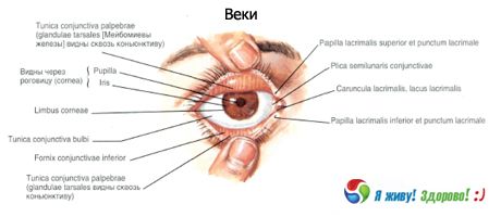
The upper eyelid (palpebra superior) and lower eyelid (palpebra inferior) are structures that lie in front of the eyeball and cover it from above and below, and completely cover it when the eyelids are closed. At the level of the edge of the orbit, the skin of the eyelids passes into the skin of the adjacent areas of the face. At the border of the upper eyelid and forehead, a transversely oriented skin ridge covered with hair protrudes - the eyebrow (supercilium). The anterior surface of the eyelid (facies anterior palpebrae) is convex, covered with thin skin with short vellus hairs, sebaceous and sweat glands. The posterior surface of the eyelid (facies posterior palpebrae) is concave, facing the eyeball. This surface of the eyelid is covered with the conjunctiva (tunica conjunctiva).
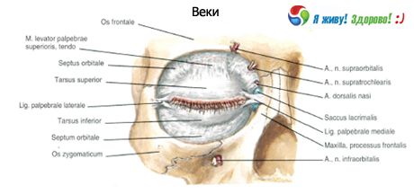
In the thickness of the upper and lower eyelids there is a connective tissue plate, the density of which resembles cartilage. This is the upper cartilage of the eyelid (tarsus superior) and the lower cartilage of the eyelid (tarsus inferior). The eyelid part of the orbicularis oculi muscle is also located here. From the upper and lower cartilages of the eyelids to the anterior and posterior lacrimal crests goes the medial ligament of the eyelid (ligamentum palpebrale mediate), common to these cartilages, which covers the lacrimal sac from the front and back. From the cartilages to the lateral wall of the orbit follows the lateral ligament of the eyelid (ligamentum palpebrale laterale), which corresponds to the lateral suture of the eyelids (raphe palpebralis lateralis).
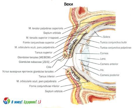
A thin, wide tendon of the muscle that lifts the upper eyelid is attached to the upper edge and the anterior surface of the cartilage of the upper eyelid. The free edge of the eyelid, limited by its posterior and anterior surfaces, respectively forms the anterior and posterior edges of the eyelids (limbi palpebrales anterior et posterior) and bears hairs located closer to the anterior edge in 2-3 rows - eyelashes (cilia).
Closer to the posterior edge of the eyelids, the openings of the modified sebaceous (meibomian) glands of the eyelid cartilage (glandulae tarsales) open, the initial parts of which are located inside the cartilaginous plate of the eyelid. There are more of these glands in the thickness of the upper eyelid (30-40) than in the lower (20-30). The edges of the upper and lower eyelids limit the transverse palpebral fissure (rima palpebrarum), which is closed on the medial and lateral sides by adhesions of the eyelids - the medial and lateral commissures of the eyelids (commissure palpebralis medialis et commissura palpebralis lateralis).
 [
[ What do need to examine?
How to examine?