Medical expert of the article

New publications
Examination of the eye under lateral (focal) and transmitted illumination
Last reviewed: 06.07.2025

All iLive content is medically reviewed or fact checked to ensure as much factual accuracy as possible.
We have strict sourcing guidelines and only link to reputable media sites, academic research institutions and, whenever possible, medically peer reviewed studies. Note that the numbers in parentheses ([1], [2], etc.) are clickable links to these studies.
If you feel that any of our content is inaccurate, out-of-date, or otherwise questionable, please select it and press Ctrl + Enter.
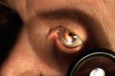
The method is designed to detect subtle changes in the anterior part of the eyeball.
The examination is conducted in a dark room using a table lamp installed to the left and in front of the patient at a distance of 40-50 cm at the level of his face. Ophthalmic loupes with a power of 13.0 or 20.0 D are used for the examination. The doctor stands opposite the patient, his feet are to the left of the latter's feet. Then the doctor takes the loupe with his right hand, slightly turns the patient's head towards the light source and directs the beam of light at the eyeball. The loupe must be placed between the light source and the patient's eye, taking into account its focal length (7-8 or 5-6 cm) so that the light rays, passing through the glass, focus on a certain area of the anterior part of the eyeball to be examined. Bright illumination of this area in contrast with the neighboring ones makes it possible to examine individual structures in detail. The method is called lateral because the loupe is located to the side of the eye.
When examining the sclera, attention is paid to its color and the state of the vascular pattern. Normally, the sclera is white, only the conjunctival vessels are visible, the marginal looped network of vessels around the cornea is not visible.
The cornea is transparent, shiny, smooth, mirror-like, spherical. Normally, the cornea does not have its own vessels. The anterior chamber of the eye is visible through the cornea, the depth of which is better seen from the side. The distance between the light reflexes on the cornea and iris determines the depth of the anterior chamber (normally, its depth in the center is 3-3.5 mm). The moisture filling the anterior chamber is normally completely transparent. In some diseases, it may contain pus, blood, flakes of exudate. When examining the iris through the cornea, note whether there are any changes in color and pattern, the presence of coarse pigment inclusions, assess the condition of the pigment border, the width and mobility of the pupil. The color of the iris depends on the amount of pigment in it and can be from light blue to dark brown. A change in the color of the iris can be detected by comparing it with the color of the iris of the other eye. In the absence of pigment, the iris is transparent, it has a red color due to the translucency of the vascular membrane (albinos). The trabecular and lacunar structure of the iris gives it an openwork appearance. The pupillary and root (ciliary) zones are clearly visible in it. A brown border is noted along the pupillary edge, which is part of the internal pigment sheet of the iris, everted onto its anterior surface. With age, this border becomes depigmented.
With lateral illumination, the pupil is defined as a black circle. The pupil can be examined using three methods: pupilloscopy, pupillometry and pupillography, but in clinical practice the first two are usually used.
A study to determine the size (width) of the pupil is usually conducted in a bright room, with the patient looking into the distance over the doctor's head. Attention is paid to the shape and position of the pupil. Normally, the pupil is round, and in pathological conditions it can be oval, scalloped, or eccentrically located. Its size varies depending on the illumination from 2.5 to 4 mm. In bright light, the pupil contracts, and in the dark, it expands. The size of the pupil depends on the patient's age, refraction, and accommodation. The width of the pupil can be measured with a millimeter ruler, or more accurately, with a pupillometer.

An important property of the pupil is its reaction to light; three types of reaction are distinguished: direct, consensual, reaction to convergence and accommodation.
To determine a direct reaction: first, both eyes are covered with palms for 30-40 seconds, and then opened one by one. In this case, the pupil of the opened eye will narrow in response to the light beam entering the eye.
The consensual reaction is checked as follows: at the moment of closing and opening one eye, I observe the reaction of the other. The study is conducted in a darkened room using light from an ophthalmoscope or slit lamp. When opening one eye, the pupil in the other eye will dilate, and when opening, it will narrow.
The pupil's reaction to convergence and accommodation is assessed as follows. The patient first looks into the distance, and then shifts his gaze to some close object (the tip of a pencil, the handle of an ophthalmoscope, etc.), located at a distance of 20-25 cm from him. In this case, the pupils of both eyes narrow.
The transparent lens is not visible when examined using the lateral illumination method. Individual areas of opacities are determined if they are located in the superficial layers: When the cataract is fully mature, the pupil becomes white.
Transmitted light study
The method is used to examine optically transparent media of the eyeball (cornea, anterior chamber fluid, lens, vitreous body ). Considering that the cornea and anterior chamber can be examined in detail with lateral (focal) illumination, this method is used mainly to examine the lens and vitreous body.
The light source is placed (in a darkened room) behind and to the left of the patient. The doctor directs the reflected beam of light into the patient's pupil using a mirror ophthalmoscope placed against his right eye. For a more detailed examination, the pupil must first be dilated with medication. When the beam of light hits the pupil, it begins to glow red, which is caused by the reflection of rays from the choroid (reflex from the fundus). According to the law of conjugate foci, some of the reflected rays enter the doctor's eye through an opening in the ophthalmoscope. If fixed or floating opacities are encountered on the path of the rays reflected from the fundus, then fixed or moving dark formations of various shapes appear against the uniform red glow of the fundus. If opacities in the cornea and anterior chamber are not detected with lateral illumination, then the formations detected in transmitted light are opacities in the lens or vitreous body. Opacities in the vitreous body are mobile, they move even when the eyeball is motionless. Cloudy areas in the lens are fixed and move only when the eyeball moves. In order to determine the depth of the opacities in the lens, the patient is asked to look up, then down. If the opacities are in the anterior layers, then in transmitted light it will move in the same direction. If the opacities are in the posterior layers, then they will shift in the opposite direction.