Medical expert of the article

New publications
Excretory urography
Last reviewed: 29.06.2025

All iLive content is medically reviewed or fact checked to ensure as much factual accuracy as possible.
We have strict sourcing guidelines and only link to reputable media sites, academic research institutions and, whenever possible, medically peer reviewed studies. Note that the numbers in parentheses ([1], [2], etc.) are clickable links to these studies.
If you feel that any of our content is inaccurate, out-of-date, or otherwise questionable, please select it and press Ctrl + Enter.
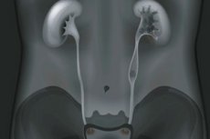
Excretory urography (or intravenous urography, IVU) is a medical procedure that is used to visualize the urinary tract using x-rays. This type of urography is performed using a contrast agent that is injected intravenously (through a vein) and filtered through the kidneys. Excretory urography is designed to evaluate the structure and function of the kidneys, ureters, ureters and bladder.
Here's how the excretory urography procedure works:
- The patient is injected with a contrast agent intravenously through a vein in the forearm or hand.
- The contrast agent circulates in the blood and passes through the kidneys.
- The kidneys filter the contrast agent from the blood and excrete it into the urine.
- A series of X-rays are then taken at different points in time after the contrast agent is injected. These images allow doctors to watch the contrast agent pass through the urinary tract and visualize it on the pictures.
Excretory urography may be used for the following purposes:
- Diagnosis of urinary tract and kidney anomalies.
- Detection of kidney and urinary tract stones.
- Bladder and urethra assessment.
- Monitoring the effectiveness of urinary disease treatment.
- Investigation of signs and symptoms such as lower back pain, blood in the urine, or frequent urination.
Excretory urography is generally considered a safe procedure, but there may be some discomfort due to the injection of contrast material. Patients may be given instructions on how to prepare for the procedure, such as prescribing restrictions on food and fluid intake in the run-up to the test.
Indications for the procedure
Excretory urography may be ordered in the following cases:
- Diagnosis of kidney and urinary tract anomalies: Excretory urography can be used to detect congenital anomalies of the structure of the kidneys, ureters, ureters and bladder.
- Suspicion of stones: The procedure may be ordered to detect the presence of stones (urolithiasis) in the kidneys or urinary tract, which may be the cause of pain and urinary problems.
- Evaluation of trauma and injury: Excretory urography can be used to evaluate the kidneys and urinary tract for suspected trauma or injury following accidents or trauma.
- Monitoring of kidney disease: Urography can be used to evaluate the kidneys and urinary tract in various kidney diseases such as glomerulonephritis, pyelonephritis or polycystic kidney disease.
- Investigation of vague symptoms: If a patient has vague symptoms related to the urinary system, such as blood in the urine, low back pain, frequent urination, or urinary incontinence, excretory urography may help establish a diagnosis.
- Surgical planning: Prior to certain surgical procedures involving the kidneys or urinary tract, excretory urography may be required for detailed assessment of organ anatomy and function.
The indications for excretory urography may vary depending on the patient's specific symptoms and clinical situation. The decision to order this procedure is usually made by the physician based on the medical history, physical examination, and other diagnostic data.
Preparation
Preparation for excretory urography may vary depending on medical practices and the requirements of the healthcare facility, but usually includes the following general steps:
- Coordinating with your doctor: Before you begin preparation, it is important to discuss the need for the test with your doctor and to make sure there are no contraindications.
- Report medical history: Tell your doctor about all of your medical conditions, allergies, and medications you are taking. This will help your doctor to take into account the specifics of your case when planning the study.
- Preparation for the contrast agent: If you are allergic to the contrast agent or have a history of allergic reactions to it, tell your doctor. Your doctor may suggest precautions such as taking antihistamines or corticosteroids before the test.
- Overnight fasting: In some cases, your doctor may recommend that you do not eat or drink (except water) after midnight before the excretory urography. This may be necessary for better visualization of the kidneys.
- Bowel cleansing: Depending on your doctor's practice and instructions, you may also need to cleanse your bowels by taking a mild laxative the evening before the exam and the morning before the procedure.
- Removal of metal jewelry: You may be asked to remove metal jewelry because it can interfere with the quality of visualization on x-rays.
- Preparation for the day of the test: Follow the instructions of your doctor and medical staff before the test. You are usually allowed to drink some water before excretory urography to excrete urine, but you should refrain from eating.
- Individualized instructions: Your individualized instructions may vary depending on the specific circumstances of your procedure and medical practice, so it is important to follow your doctor's recommendations.
Drugs used in excretory urography
This procedure uses a special contrast agent to help improve the visibility of the urinary organs on x-rays.
Drugs that may be used during excretory urography include the following:
- Contrastagents: Contrast agents such as monoiodinated contrast (MDCT), iodine contrast agents, or other agents are commonly used for excretory urography. These agents are injected into the patient's body to improve the visibility of the kidneys, ureters, bladder, and urethra on x-rays.
- Sedation drugs: In some cases, sedation or anesthesia drugs may be used to provide comfort to the patient and reduce anxiety during the procedure.
- Drugs to prevent allergic reactions: If the patient is allergic to the contrast agent, the doctor may prescribe antihistamines or corticosteroids to prevent allergic reactions.
- Bloodpressure and pulse control medications: If necessary, medications may be used to control the patient's blood pressure and pulse.
All drugs and medications used during excretory urography must be prescribed and administered by the physician or medical staff performing the procedure. The physician will take into account the patient's medical history, allergies and other factors to select the appropriate drugs and dosage, and will advise the patient of possible risks and side effects.
Contrast agents
The contrast agents used in excretory urography help visualize the urinary tract and assess its function on x-rays. There are several types of contrast agents that can be used for this procedure. The following are some of them:
- Iodine-containing contrast agents: These contrast agents contain iodine and are commonly used in excretory urography. They allow x-rays to pass easily through the organs of the urinary tract and make them visible on images. Examples of iodine-containing contrast agents include iodolipol, iodamidol, and others.
- Non-complex contrast agents: These contrast agents do not form stable chemical compounds with calcium and magnesium molecules, allowing them to be easily excreted through the kidneys into the urine. This makes them well suited for evaluating renal function. Examples of non-complex contrast agents include meglumic acid and meglumic sulfate.
- Osmolar contrast agents: These contrast agents are commonly used in older excretory urography techniques. They have a high osmolarity and may cause more minor side effects than more modern contrast agents. An example of an osmolar contrast agent is diatrizoate.
The choice of a particular contrast agent may depend on the physician's medical practice, location, and preference, as well as patient characteristics and history. The physician will usually select the contrast agent that best suits the purpose of the study and minimizes the risk of allergic reactions or side effects.
Technique of the an excretory urography
The procedure is carried out as follows:
-
Patient Preparation:
- The patient may be asked to take tests before the procedure to check kidney function and blood creatinine levels.
- The patient should be on an empty stomach or on a light diet in the run-up to the study, following the physician's instructions regarding food and fluid intake.
- Before the procedure, the patient may be asked to remove metal objects (jewelry, coins, etc.) so that they do not interfere during the x-rays.
-
Injection of a contrast agent:
- Once the patient is in the radiology room, the medical staff will insert an intravenous catheter into a vein on the forearm or other location.
- A contrast agent is injected through this catheter. The doctor monitors the process of spreading the contrast agent through the kidneys and urinary tract.
-
Obtaining X-rays:
- After the contrast agent is injected, the patient is given a series of x-rays at different points in time.
- Images are taken as the contrast agent passes through the kidneys, ureters, and urethra. This allows the structure and function of the urinary tract to be visualized.
-
Completion of the procedure:
- After the x-rays are completed, the catheter is removed.
- The patient can return to normal activities after the procedure if no complications arise.
The time required to perform excretory urography is usually several hours, including preparation and performance of the procedure. The results are evaluated by a radiologist who will draw conclusions about the condition of the urinary tract and issue a report that will be shared with the patient's doctor.
Types of excretory urography
Depending on the specific objectives and areas to be examined, there are several different types of excretory urography. Here are some of them:
- Intravenous pyelography (IVP): This is the most common type of excretory urography. During IVP, a contrast agent is injected into a vein and subsequent x-rays are taken at different time intervals. This method evaluates the kidneys, ureters, and bladder.
- Retrograde pyelography: This method is used to examine the ureters and renal pelvis in more detail. A contrast agent is injected through a catheter inserted into the bladder through the urethra. X-rays are then taken.
- Ureteropyelography: This method evaluates the condition of theureters. A contrast agent is injected directly into the ureters through a catheter. X-rays are then taken to study the anatomy and patency of the ureters.
- Pediatric excretory urography: This type of excretory urography is designed to examine the urinary system in children. The procedure is adapted to the age and size of the child.
- Positive ContrastPyelography: This method uses positive contrast agents that appear white on X-rays. They allow you to see the contours of the urinary system more clearly.
- Negative ContrastPyelography: This uses negative contrast agents that appear black on x-rays. This method can be useful for detecting some abnormalities.
The choice of excretory urography depends on the specific clinical questions and goals of the study, as well as the age and condition of the patient. These procedures can help doctors identify abnormalities, infections, stones and other problems in the urinary system and develop an appropriate treatment plan.
Excretory urography in children
Excretory urography may also be performed in children to evaluate the urinary system. This procedure can be especially helpful in detecting abnormalities, infections, stones, or other problems in the urinary system in children. Here are some features of excretory urography in children:
- Age: The procedure can be performed in both newborns and older children. The age of the child affects the specifics and approach of the study.
- Preparation: Preparation for excretory urography in children may include the same elements as in adults, such as fasting before the procedure and taking contrast agent. However, the preparation should be adapted to the child's age and condition.
- Contrastagent: The contrast agent used in the study should be adjusted for the child's age and weight. The dosage of contrast may vary depending on the child's age.
- X-rays: X-rays of the urinary tract are performed for children using a contrast agent. The X-ray machine and cine screen are adapted to ensure the safety and comfort of children.
- Special Considerations: Children may require special considerations such as anesthesia or sedation to make the procedure less stressful and painful.
- Supervising Actions: Doctors and medical staff are required to monitor children more closely during the procedure to ensure their safety and comfort. Parents may be present during the study to support the child.
Excretory urography in children can be used for a variety of purposes, including detecting urinary tract abnormalities, evaluating the cause of low back pain, detecting infections, or determining the presence of stones in the urinary system. If necessary, doctors may recommend this procedure to get more information about your child's health.
Contraindications to the procedure
Contraindications can vary depending on the specific circumstances and condition of the patient, but here are some general contraindications to excretory urography:
- Allergy to contrast agent: If the patient has a known allergy to the contrast agent used for excretory urography, this may be a contraindication. The physician should consider alternative methods of examination or take precautions such as pre-treatment with antihistamines or corticosteroids.
- Severe renal impairment: Patients withsevere renal impairment or chronic renal failure may have problems with excretion of contrast medium. In such cases, urography may be dangerous and not applicable.
- Pregnancy: X-rays may be contraindicated during pregnancy due to potential risk to the fetus. If pregnancy is a possibility, the physician should consider alternative diagnostic methods or postpone the study until a safer time.
- Renalcolic or acute renal failure: In acute renal colic or severe renal failure, urography may be contraindicated because of the risk of additional renal damage or worsening of the condition.
- Patients with asthma or other allergic reactions: Patients with allergic reactions to medications may require special precautions such as prior administration of antihistamines or corticosteroids.
- Children and elderly patients: Children and elderly patients may have special risks and limitations of excretory urography and the decision to perform the study should be evaluated individually.
Normal performance
Normal values for excretory urography may vary depending on the age, sex and individual characteristics of the patient. They also depend on which parts of the urinary system are being evaluated as part of the study. Here are some of the common normal values that can be evaluated with excretory urography:
- Passage of the contrast agent: The contrast agent must pass through the ureters and into the bladder. This normally occurs at certain time intervals after the contrast is injected.
- Bladder filling: The bladder should be completely filled with contrast agent.
- Anatomydefinition: Doctors evaluate the anatomy of the urinary system on x-rays. Normal anatomical structures should be clear and without abnormalities.
- Urinary tract clearance: Doctors may assess for narrowings (strictures) or other obstructions in the urinary tract that could make it difficult to pass urine.
- Ruling out the presence of stones: Excretory urography may be useful to detect the presence of stones (stones) in the urinary system.
When interpreting the results of excretory urography, it is important to consider the patient's medical history, symptoms, and clinical findings. Normal values may vary, and even small abnormalities or abnormalities may have different clinical significance. The final judgment and interpretation of the results should always be provided by the physician who performed the study and has all the necessary information about the patient's condition.
Complications after the procedure
Excretory urography is generally considered a relatively safe procedure, but as with any medical test, certain complications and side effects can occur. Here are some of the potential complications:
- Allergic reaction to contrast agent: Some patients may have an allergic reaction to the contrast agent injected during the procedure. This may manifest as itching, skin rash, redness, swelling, or even more serious allergic reactions. Patients who are allergic to the contrast agent should inform their doctor before the procedure.
- Acute kidney injury: Rarely, but occasionally, contrast agent may adversely affect renal function and cause acute kidney injury, especially in patients with pre-existing kidney problems.
- Unpleasant sensations: The patient may experience discomfort or burning sensations while the contrast agent is being injected through a catheter or vein.
- Swelling or pain at the injection site: The site where the catheter or contrast agent was injected can sometimes be painful or cause a small amount of swelling.
- Ionizing radiation: Excretory urography involves the use of X-rays, which may increase health risks with prolonged and repeated use.
- Other complications: Although rare, other complications such as infections or bleeding can occur, especially if the procedure is not performed correctly.
It is important to note that the risk of complications after excretory urography is usually low, and many patients successfully undergo this procedure without any problems.
Care after the procedure
After an excretory urography procedure, some care and monitoring of your condition may be necessary. Here are some general guidelines for care after excretory urography:
- Rest: You may be advised to spend some time resting after the procedure. Relax and allow yourself to recover.
- Hydration: After excretory urography, it is important to drink enough water to help the body eliminate the contrast agent from the urinary system. Drinking water can also help prevent kidney stones from forming.
- Urination: It is important to urinate regularly after the procedure. This will help remove the contrast agent from the urinary tract. Do not hold your urine if the need arises.
- Monitor yourcondition: After excretory urography, look out for any unusual symptoms or complications such as allergic reactions, swelling, rash, pain, or anxiety. If you experience any of these symptoms, contact your doctor.
- Diet: You may be advised to follow a certain diet or limit certain foods for a period of time after the procedure. Follow your doctor's recommendations on this matter.
- Avoid physical activity: You may be advised to avoid strenuous physical activity and heavy lifting for a few days after the study to avoid injury to your urinary system.
- Keep track of your medications: If you have been prescribed any medications after the procedure, follow your doctor's instructions about taking them.
- Follow yourdoctor's recommendations: It is important to follow all the recommendations and instructions your doctor will give you after the procedure.
Review urography and excretory urography
These are two different types of x-rays that are used to visualize the urinary tract and assess its function. Here are their main differences:
-
Review urography:
- Review urography is also known as standard urography or proximal urography.
- In review urography, the patient is injected with a contrast agent intramuscularly or intravenously.
- After contrast is injected, X-rays are taken within a few minutes. The images show the structures of the urinary tract, including the kidneys, ureters, and upper parts of the ureters.
- This study is commonly used to evaluate the anatomy of the urinary tract and to detect abnormalities, stones, or tumors in the upper parts of the urinary system.
-
Excretory urography:
- Excretory urography (intravenous urography, IVU) also involves injecting a contrast agent into the patient, but intravenously through a vein in the forearm or arm.
- An important characteristic of excretory urography is the instantaneous image. After contrast is injected, X-rays are taken at different points in time to track how the contrast agent passes through the kidneys, ureters, ureters and finally the bladder.
- Excretory urography is widely used to evaluate kidney function and diagnose various diseases of the urinary system, such as stones, tumors, strictures (narrowings) and other pathologies.
Both types of urography can provide important information about the urinary tract, but the choice depends on the clinical situation and the goals of the study. Doctors choose the appropriate method depending on the symptoms, medical history, and specific questions that need to be addressed by urography.