Medical expert of the article

New publications
Endophthalmitis in children
Last reviewed: 04.07.2025

All iLive content is medically reviewed or fact checked to ensure as much factual accuracy as possible.
We have strict sourcing guidelines and only link to reputable media sites, academic research institutions and, whenever possible, medically peer reviewed studies. Note that the numbers in parentheses ([1], [2], etc.) are clickable links to these studies.
If you feel that any of our content is inaccurate, out-of-date, or otherwise questionable, please select it and press Ctrl + Enter.
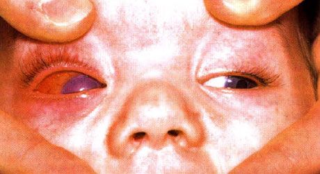
Endophthalmitis develops when the infectious process is localized in the cavity of the eyeball. The term panophthalmitis is used when the infection spreads progressively, affecting all tissues of the eye. Diagnosis of endophthalmitis in children presents certain difficulties, which is associated with the complexity of conducting research. Usually, in the presence of this disease, one can detect:
- such an etiological factor as trauma, surgery, etc.;
- swelling of the eyelids;
- conjunctival injection and chemosis;
- uveitis;
- hypopyon;
- dilation of retinal vessels.
The severity and malignancy of the clinical course of endophthalmitis depend on the route of infection and the type of pathogen. For example, Streptococcus spp. or Pseudomonas cause rapidly progressing endophthalmitis with a severe clinical course. Endophthalmitis caused by Staphylococcus spp., especially Staph, epidermidis, is characterized by a late onset and a relatively benign course. Fungal endophthalmitis, as a rule, is relatively mild, but the development of complications cannot be ruled out.
 [ 1 ], [ 2 ], [ 3 ], [ 4 ], [ 5 ], [ 6 ], [ 7 ], [ 8 ], [ 9 ], [ 10 ], [ 11 ], [ 12 ]
Cause of endophthalmitis in children
- Trauma: surgical intervention; penetrating wound.
- Keratitis: the pathogen penetrates through Descemet's membrane, causing infectious anterior uveitis, which creates conditions for the development of endophthalmitis.
- Metastatic endophthalmitis against the background of meningitis (especially meningococcal), infective endocarditis and otitis media, as well as general infection. In many cases, endophthalmitis is bilateral and is often diagnosed late due to the extreme significance of the underlying disease.
Possible infectious agents
Bacterial flora
Most often, endophthalmitis, especially postoperative, is caused by Streptococcus and Staphylococcus spp. Posttraumatic endophthalmitis is usually provoked by Proteus and Pseudomonas, often in combination with other bacterial flora. In the presence of Pseudomonas, specific keratitis develops.
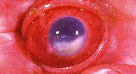
Hypopyon accompanying endophthalmitis. The background was keratitis caused by non-closure of the palpebral fissure. Although the eye was saved due to timely administration of antibacterial therapy, visual acuity remained low after 5 years due to the development of amblyopia
Fungal flora
The infectious process caused by Candida spp. usually accompanies immunodeficiency or, in other words, more often affects children with severe somatic pathology.
Research
- Gram staining of smears.
- Staining of smears according to Giemsa, especially to exclude fungal flora.
- Blood culture for sterility.
- Diagnostic puncture of the anterior chamber and/or vitreous body followed by bacteriological examination.
Samples should be immediately inoculated onto a Petri dish with blood agar, thioglycollate medium and "chocolate" agar. For detection of fungal flora, cultivation in Sabouraud nutrient medium and blood agar is used.
To clarify the degree of involvement of the posterior segment of the eye in the pathological process in case of disease of the anterior segment, an ultrasound examination is performed. A general examination helps to exclude the metastatic nature of endophthalmitis.
Where does it hurt?
Other forms of endophthalmitis
The course of toxocariasis and toxoplasmosis sometimes resembles the clinical picture of endophthalmitis. In Behcet's disease, uveitis is so severe that it imitates endophthalmitis.
Infectious conjunctivitis
The diagnosis of conjunctivitis is based on the following clinical signs:
- mucopurulent discharge;
- conjunctival injection, accompanied in some cases by hemorrhage and swelling;
- lacrimation;
- feeling of discomfort in the eye;
- mild itching that is not a pathognomonic symptom;
- vision does not deteriorate, although the patient may be bothered by a feeling of “fog” before the eyes, which is associated with a large amount of mucous discharge;
- a feeling of "sand" in the eyes, especially in cases of concomitant keratitis.
Diagnosis
- The diagnosis is established on the basis of the medical history, examination of discharge from the conjunctival cavity, and the presence of a corresponding general disorder (inflammatory process of the upper respiratory tract, etc.)
- Research:
- visual acuity testing - decreased vision is usually associated with the presence of abundant mucopurulent discharge or concomitant keratitis;
- Slit lamp examination reveals changes in the conjunctiva and, in some cases, combined keratitis;
- assessment of the cleanliness of the skin (to exclude rash) and the condition of the mucous membranes.
- Laboratory research.
Most pediatricians and ophthalmologists do not perform laboratory diagnostics when a patient first comes to them. Since conjunctivitis
is very common, and the viral or bacterial agents that cause it do not pose a serious threat and are easily treated with adequate antiviral and antibacterial therapy, there is no need to perform a culture. Culture is indicated in cases of severe clinical course, chronic and recurrent (after discontinuing antibiotics) processes, as well as follicular and atypical forms of the disease.
What do need to examine?
How to examine?
Who to contact?
Treatment of endophthalmitis in children
Antibacterial therapy
Bacterial endophthalmitis. Specific antibacterial treatment is prescribed based on the individual sensitivity of the microbial flora, identified by sowing on various media. If the sensitivity of the microflora is unknown, the following drug regimens are recommended:
- Installations:
- instillations of gentamicin solution (preferably preservative-free) every hour;
- instillations of 5% cefuroxime solution (preferably preservative-free) every hour;
- instillations of 1% atropine solution (for children under 6 months of age, 0.5% atropine is instilled) twice a day.
- Subconjunctival injections (if vitreous puncture is necessary, subconjunctival injections are combined with surgical intervention):
- gentamicin - 40 mg;
- cefazolin - 125 mg.
- Intravitreal injections:
- gentamicin (diluted 0.1 mg in 0.1 ml);
- ceftazidime (diluted 2.25 mg in 0.1 ml).
- General uses of antibiotics:
- gentamicin - intravenously, at a daily dose of 2 mg/kg of weight;
- cefuroxime - intravenously, at a daily dose of 60 mg/kg of weight, in several doses.
Endophthalmitis of flexible etiology. When Candida fungi are isolated, ketoconazole or amphotericin B in combination with flucytosine are usually prescribed. Most other representatives of the fungal flora are sensitive to amphotericin B, which is administered intravitreally (5 mcg).
Vitrectomy
In some cases, early vitrectomy may play a role in order to completely sanitize the infectious focus, as well as remove foreign bodies and necrotic tissue. An antibiotic is administered intravitreally and subconjunctivally at the same time as vitrectomy.