Medical expert of the article

New publications
Electromyography of pelvic floor and bladder muscles
Last reviewed: 07.07.2025

All iLive content is medically reviewed or fact checked to ensure as much factual accuracy as possible.
We have strict sourcing guidelines and only link to reputable media sites, academic research institutions and, whenever possible, medically peer reviewed studies. Note that the numbers in parentheses ([1], [2], etc.) are clickable links to these studies.
If you feel that any of our content is inaccurate, out-of-date, or otherwise questionable, please select it and press Ctrl + Enter.
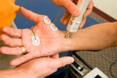
Electromyography is the recording of the bioelectric potential of a muscle.
In urodynamic studies, EMG is used to record the activity of the pelvic floor striated muscles: the pubococcygeus muscle (M. pubococcygeus), the levator ani muscle (m.levator ani), and the anal sphincter (rabdosphincter). Electromyography data are necessary to create a complete picture of the urination cycle: during urine accumulation, the muscles prevent it from flowing out, and during emptying, they relax, ensuring adequate emptying. During bladder contraction, the muscles should not only relax, but do so in a coordinated manner, without delay. Urodynamic studies supplemented by EMG allow recording the level of bioelectrical activity of the pelvic floor muscles during filling and emptying.Video urodynamics can be used to assess the function of smooth muscles (e.g., the neck of the bladder).
Technically, EMG is a study of electrical potentials generated by depolarization of striated muscle. This is the result of the work of the motor neuron and the muscle it innervates. Recording is performed using cutaneous or needle electrodes. Urodynamic research is more conveniently combined with the use of cutaneous electrodes that collect information from a group of muscles located directly beneath them. A needle electrode can be placed directly into the muscle and record a separate EMG potential. Needle electrodes can be concentric, monopolar, and bipolar. They are often used to conduct additional studies logically related to urodynamic studies, but separated from them in time. Data interpretation is performed jointly. Experts also classify the following neurophysiological methods as urodynamic studies:
- study of nerve conduction along the n. pudendus;
- study of the bulbocavernous reflex;
- somatosensory evoked potentials (spinal and cortical).
They are recorded using both stationary and portable equipment.
 [
[ What do need to examine?
What tests are needed?