Medical expert of the article

New publications
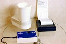
Cystometry
Last reviewed: 07.07.2025

All iLive content is medically reviewed or fact checked to ensure as much factual accuracy as possible.
We have strict sourcing guidelines and only link to reputable media sites, academic research institutions and, whenever possible, medically peer reviewed studies. Note that the numbers in parentheses ([1], [2], etc.) are clickable links to these studies.
If you feel that any of our content is inaccurate, out-of-date, or otherwise questionable, please select it and press Ctrl + Enter.
Cystometry is a basic method of urodynamic studies, during which both phases of the micturition cycle are examined - filling (accumulation) and emptying, and the dependence of intravesical pressure on the degree of filling of the bladder is studied. Cystometry allows assessing the function of the detrusor and urethra in different periods. Thus, normally, in the filling phase, the bladder does not contract and is passive, and the urethra is closed (contracted). In the emptying phase, the bladder contracts, and the urethra relaxes, which ensures a normal flow of urine. Filling is assessed in terms of sensitivity, capacity, stability of compliance and competence: that is, both the motor and sensory components of the micturition reflex are examined.
Cystometry is an invasive examination. Before performing it, the patient's medical history is studied, a physical examination is performed, and the urination diary and results of a general urine analysis are assessed. The physical examination can be called neurourological and urogynecological in terms of specificity. Some reflexes (anal, bulbocavernous) and cognitive function are determined. For women, a vaginal examination, assessment of the pelvic floor muscles, and, if indicated, a Q-tip or straight catheter test to determine the mobility of the urethra, a test with pads are required. For men, a digital rectal examination is required, and, if necessary, an ultrasound examination (ultrasound) of the prostate.
Indications for cystometry
- pollakiuria,
- nocturia,
- urgency of urination,
- enuresis,
- difficulty in "starting" urination,
- urinary incontinence,
- the presence of residual urine in the bladder (retention),
- dysuria in the absence of an inflammatory process in the urinary system.
Main evaluation criteria for cystometry
Criterion |
Characteristic |
Sensitivity |
A subjective sensation that occurs when the bladder is full. It is defined from the moment of the first feeling of filling until a strong urge |
"Stability" (in old terminology) or absence of involuntary contractions of the detrusor |
During the filling phase, the bladder is inhibited and does not contract. Micturition begins with a voluntary contraction of the detrusor |
Compliance |
The ability of the urinary bladder to maintain low intraluminal pressure at different volumes of its filling. Determined by the formula C=V/P of the detrusor (ml/cm H2O) |
Capacity |
Cystometric - the volume of the bladder at which the command to urinate is given. Maximum cystometric - the volume at which the patient can no longer hold back the urge to urinate. |
Competence (of the urethra) |
The ability to maintain and, if necessary, increase the pressure in the closure zone, ensuring the constancy of the difference between the urethral and bladder pressure in its favor (ensuring urine retention during filling) |
Cystometry can be simple single-channel, when only intravesical pressure is recorded. Such a study is carried out in two modes: intermittent, when filling the bladder with a sterile solution/water alternates with periods of pressure recording (a single-channel catheter is used), or continuous, when filling and recording are carried out simultaneously (a two-channel catheter is used).
Currently, dual-channel cystometry is considered standard, when intravesical and intra-abdominal pressures are recorded simultaneously. A dual-channel catheter is used to measure intravesical pressure (usually 6-10 CH) and a rectal balloon catheter is used to measure intra-abdominal pressure.
Catheters filled with water, air, and "micro-type" catheters with a piezoelectric sensor at the end can be used. Water catheters are the most accessible and widely used. In the future, it is possible to switch to air or "micro-type" catheters, which provide more accurate measurements free from the influence of the hydrostatic component. Catheters are connected to pressure sensors and a computer system that records the readings. The study is carried out in a standing, sitting, or lying position. Pressure sensors must be placed at the level of the pubic symphysis. In expert-class laboratories, the number of measurement channels is sometimes increased to six, combining cystometry with EMG and constant X-ray control (video urodynamic study).
The International Continence Society (ISC) recommends a minimum list of requirements for equipment for cystometry:
- two pressure measurement channels with display and safe storage of three pressure readings (bladder, abdominal, detrusor);
- one channel for measuring urine flow with display and storage of information;
- recording of indicators of the volume of introduced and the volume of excreted urine (in graphic and digital form);
- adequate scales and measurement scales without loss of information outside the scale boundaries;
- accounting of standard information recording.
Methodology for performing cystometry
The examination begins with the patient being placed in a chair or on a couch for processing the "field", installing catheters, connecting them to sensors, and checking the adequacy of their operation. The bladder must be empty. During inpatient urodynamics, filling is carried out at a rate of 10-100 ml/min (depending on the patient's age and bladder capacity). Outpatient urodynamic examination involves natural filling of the bladder. The filling volume is calculated according to the capacity: for adults - 400-500 ml. for children - according to the formula 30 + 30p, where p is the patient's age in years.
During filling, the patient's sensations, pressure and volume indicators are recorded. The main parameters recorded during urination (voiding cystometry) are pressure, flow rate and volume. During the study, the main events are marked on the graph:
- cough to confirm that the pressure transmission is OK (carried out at the beginning, at the end and after every 100 ml of filling):
- start of infusion;
- first sensation;
- first urge to urinate;
- normal urge to urinate;
- strong urge to urinate;
- spontaneous and cough- or straining-induced urine leakage;
- maximum cystometric capacity;
- stop the infusion and start urinating;
- non-specific sensations, pain, urgency;
- artifacts (possibly with comments).
In the research report, all events must be detailed by pressure readings of all recording channels and filling volume at the time of the event.
Decoding the results
Urodynamic disturbances determined by cystometry:
- increased sensitivity - the occurrence in the early stages of filling of the first sensation or urge, a strong prolonged urge to urinate;
- decreased sensitivity
- decreased sensitivity during filling;
- lack of sensitivity - there is no sensitivity during the entire filling phase of the bladder;
- decreased compliance - impaired ability to maintain low intravesical pressure during filling, which leads to a decrease in cystometric capacity;
- detrusor overactivity - involuntary increases in detrusor pressure of varying amplitude. It can be neurogenic (neurological cause) and idiopathic. Neurogenic detrusor overactivity is characterized by a higher amplitude of contractions,
- urinary incontinence due to detrusor overactivity (urge urinary incontinence):
- stress urinary incontinence: loss of urine due to increased abdominal/intra-abdominal pressure:
- IVO - an increase in the detrusor micturition pressure and a decrease in the flow rate when they are recorded synchronously (standardized only for men; for women, clear criteria have not yet been defined). IVO is often caused by an enlarged prostate in men and pelvic organ prolapse in women (see "Pressure/Flow Ratio Study");
- dysfunctional urination (pseudo dyssynergia) uncoordinated relaxation of the pelvic floor muscles and contraction of the detrusor during urination in the absence of a neurological disorder, which leads to impaired emptying of the bladder. To diagnose this disorder, cystometry is combined with EMG of the pelvic floor muscles;
- detrusor-sphincter dyssynergia - a contraction of the urethra and periurethral striated muscles, competitive with the contraction of the detrusor, recorded during voiding. In this case, the flow of urine may be interrupted. It is determined only in patients with spinal cord injuries. To diagnose detrusor-sphincter dyssynergia, cystometry is supplemented with EMG and/or performed as part of a videourodynamic examination.
Thus, cystometry is of great clinical importance, as it helps to correctly interpret the symptoms of urination disorders and choose the most effective type of treatment.
 [ 1 ], [ 2 ], [ 3 ], [ 4 ], [ 5 ]
Pressure/flow relationship study
It consists of measuring intravesical pressure, intra-abdominal pressure and volume flow rate during the entire urination phase. The study is used to analyze voiding disorders and determine their cause (either IVO or bladder contractility disorder).
From the point of view of urination physiology, it is believed that the urine flow picks up speed when the detrusor pressure begins to exceed the urethral pressure. This value is called the opening pressure of the urethra (P det, open). Subsequently, the flow rate reaches its maximum (Qmax), which is determined by the ratio between the detrusor and urethral pressures. As soon as the detrusor pressure ceases to exceed the pressure in the urethra, the bladder is no longer able to expel urine, and the flow rate becomes zero.
Complete emptying of the bladder is ensured by three components:
- sufficient in amplitude and duration of detrusor contraction;
- adequate and timely reduction of urethral resistance (opening of the sphincter);
- absence of mechanical obstruction.
Additionally, to assess the coordination of the pelvic floor muscles and detrusor contractions, EMG can be performed, and, according to special indications, video urodynamic examination.
The flow/volume ratio study is performed after filling cystometry, when the patient expresses the desire to urinate and the bladder stops filling. The recommended catheter size is 7-8 CH, so as not to create additional obstruction to the urine flow. The uroflowmeter is placed as close as possible to the external opening of the urethra to record the flow without artificial delay. The study is performed in the most comfortable conditions, without external irritants and provocations. The following recorded indicators are used for interpretation:
- intravesical pressure - Pves (mm H2O);
- abdominal/intra-abdominal pressure - Рabd (mm H2O);
- detrusor pressure - Pdet (mm H2O)
- maximum detrusor pressure (cm H2O);
- detrusor pressure at maximum flow (cm H2O);
- residual urine volume.
Flow/volume ratio testing is the only way to differentiate men with low Qmax due to detrusor dysfunction from patients with true IVO. IVO is indicated by low Qmax values with high intravesical pressure. On the other hand, the combination of low intravesical pressure with relatively high Qmax values indicates non-obstructive urination. Patients with low intravesical pressure and Qmax values may be suspected of having detrusor dysfunction, either primary or due to IVO.
For the convenience of assessing the parameters of obstruction and contractility, a large number of nomograms have been proposed. Two of them are most often used.
Abrams-Griffiths nomogram (1979). To construct it, the authors used pressure/flow ratio graphs to identify patients with IVO. The nomogram allows urination to be defined as obstructive (high pressure, low flow), non-obstructive (low pressure and high flow), or ambiguous. The boundaries between the three zones of the nomogram were determined empirically.
The Schafer nomogram (1985) is an alternative method for interpreting the degree of obstruction. The author used the same basic principles as in creating the Abrams-Griffiths nomogram. The pressure/flow ratio was estimated taking into account the concept of elasticity and distensibility of the urethra. The analysis allowed us to introduce the concept of "passive urethral resistance", which quantitatively interprets the pressure/flow study data. Passive urethral resistance is defined as the ratio of the minimum value of the opening pressure of the urethra and the constant C. These parameters reflect the optimal conditions for urine outflow from the bladder for a given act of urination with a relaxed state of the urethra and the lowest possible urethral resistance. The location of the graph and the shape of the loop of the linear ratio of passive resistance of the urethra depend on the nature and degree of obstruction. By transferring the simplified pressure/flow study graph to the nomogram, it became possible to evaluate the degree of obstruction on a 7-point scale (from 0 to VI). Comparison of the proposed methods in the clinical evaluation of obstruction showed their complete coincidence, which proves the validity of the underlying theoretical assumptions.
The urine flow/volume ratio is standardized only for men, for whom nomograms have been developed to assess micturition function. Approaches to assessing obstruction in women are under development. The following urodynamic criteria are currently used to determine female obstruction: Pdet/Qmax >35 cm H2O with Qmax <15 ml/s.
When examining men, the urine flow/volume ratio is the "gold standard". Timely determination of the nature of urodynamic disorders (primarily IVO) is of practical importance in the treatment of patients with prostate adenoma, since without taking this factor into account, the functional results of surgical treatment are significantly worsened. It is believed that about 25-30% of patients referred for surgery based on the results of a comprehensive examination meet the urodynamic criteria for obstruction associated with prostate disease, and up to 30% of patients with reduced detrusor contractility without signs of obstruction undergo surgical treatment.
Currently, the European Association of Urologists has developed strict indications for conducting flow/volume studies in patients who are scheduled for surgery for prostate adenoma:
- age less than 50 years;
- age over 80 years;
- residual urine volume more than 300 ml;
- Qmax >15 ml/s;
- suspected neurogenic dysfunction;
- previous radical surgery on the pelvic organs;
- in case of unsatisfactory results of previous surgical treatment
It is proposed to add an additional item to the list of indications - discrepancy between the level of complaints (using the international system of total assessment of prostate symptoms (IPSS)] and the data of the primary uroflowmetric screening (pronounced complaints and minor urination disorders or minor complaints with pronounced urination disorders determined by uroflowmetry).
Combined urodynamic testing is also recommended for patients with concomitant diabetes mellitus before planned surgical or minimally invasive treatment. Timely flow/volume testing significantly improves surgical treatment results, avoids diagnostic errors, and thus improves patients' quality of life.
Leak Point Pressure Study
Conducted in patients with insufficiency of the urethral locking function for various reasons. A distinction is made between abdominal and detrusor pressure at the leak point. Abdominal pressure is measured during coughing or straining. It is preferable to measure during straining, since it is necessary to determine the minimum pressure that leads to leakage. During a cough test, the amplitude is usually higher than the minimum required. The most important parameter is detrusor pressure, when urine leakage occurs due to an increase in detrusor pressure without a “stress” provocation or straining. The intravesical pressure measured at the beginning of urination/leakage is defined as the opening pressure.
In patients with IVO, this indicator is quite high. In some cases, during obstruction, the detrusor pressure exceeds 80 cm H2O (one of the IVO indicators). In this situation, this is a reflection of urethral resistance, and not a characteristic of the continence function. Patients with pathologically high detrusor leakage may simultaneously have a low abdominal pressure indicator. Men with damage to the striated sphincter (for example, after radical prostatectomy) have a low detrusor pressure at the leak point, as do healthy women with a short, easily opening urethra. Thus, it is difficult to judge the function of the detrusor itself by this indicator.
The clinical meaning of determining the detrusor pressure at the leak point is to predict the situation in the upper urinary tract in the presence of simultaneous obstruction (usually functional) and urinary incontinence in patients with neurogenic urination disorders. In such patients, bladder compliance decreases, high-amplitude detrusor overactivity is diagnosed, which leads to the development of retrograde hydraulic pressure and damage to the upper urinary tract. Values exceeding 40 cm H2O are considered critical. For this group of patients, it is appropriate to measure the detrusor leak pressure as part of a video-urodynamic study.
Abdominal leak pressure is used primarily to diagnose stress urinary incontinence in women:
- type III is characterized by pressure below 80 cm H2O (due to insufficiency of the internal sphincter);
- for type II - above 80 cm H2O (due to hypermobility of the urethra).
Standard equipment, any type of catheter (water, air-filled, "microtype") of the smallest possible size for measuring intravesical pressure and a standard rectal catheter are used for the study. When interpreting the data, it is important to correctly calculate the parameters taking into account the patient's position, starting pressure and possible artifacts.
Intraurethral pressure profile
It is a measurement and graphic display of intraluminal pressure along the entire length of the urethra. There are two main methods of measurement: static and dynamic. For static measurement, the theoretical basis is the position that the pressure of the urine flow should be the force that is necessary to open the urethra and begin urination. Thus, the pressure/resistance is measured at each point along the entire length of the urethra. During static passive profilometry, the patient is at rest. During stress profilometry, the patient is asked to cough and strain periodically, during which the urethral resistance is measured.
Dynamic measurement of the intraurethral pressure profile is performed at the moment of urination. Measured parameters:
- urethral closure pressure - the difference between urethral and bladder pressures;
- urethral closure pressure (stress) - the difference between urethral and bladder pressures during coughing;
- maximum urethral pressure - the maximum recorded pressure in the measurement zone;
- maximum urethral closure pressure - the pressure at the point where urethral pressure most exceeds bladder pressure;
- maximum urethral closure pressure (stress) - the pressure at the point where urethral pressure most exceeds bladder pressure during coughing;
- urethral closure pressure profile the difference between urethral and bladder pressures at all points along the urethra during coughing. Positive peaks correspond to urine retention zones (the pressure in the urethra is higher than the bladder pressure), and negative peaks correspond to incontinence zones (the bladder pressure is higher than the urethral pressure);
- functional profile length is the length of the urethra where the urethral pressure is higher than the bladder pressure;
- transmission of pressure - is determined by the ratio of the increase in intravesical pressure to the increase in urethral pressure during coughing, expressed as a percentage. Normally, the ratio is 1:1 (100%). With hypermobility of the urethra, when its proximal part loses its normal intra-abdominal position and is outside the transmission zone, the indicator decreases.
The intraurethral pressure profile is studied using standard equipment with a three-way catheter with channels for infusion, measurement of intravesical and urethral pressure. A microtype catheter is preferred. A special device, a puller, is used to advance the catheter along the urethra at a constant speed and fix it at the external opening.
The study of the intraurethral pressure profile is included in the standard examination of women suffering from urinary incontinence. Less often it is performed on men (mainly in case of decompensation of the external sphincter and postoperative urinary incontinence).
There is no unanimous opinion on the study of the intraurethral pressure profile to determine urodynamics. Different specialists prefer one or another method of measuring it, and some refuse to conduct it at all. Nevertheless, in a number of clinical situations, this study is necessary and allows for an assessment of the urodynamic situation as a whole, and therefore more accurately.
What do need to examine?
What tests are needed?