Medical expert of the article

New publications
Elastic pseudoxanthoma: causes, symptoms, diagnosis, treatment
Last reviewed: 07.07.2025

All iLive content is medically reviewed or fact checked to ensure as much factual accuracy as possible.
We have strict sourcing guidelines and only link to reputable media sites, academic research institutions and, whenever possible, medically peer reviewed studies. Note that the numbers in parentheses ([1], [2], etc.) are clickable links to these studies.
If you feel that any of our content is inaccurate, out-of-date, or otherwise questionable, please select it and press Ctrl + Enter.
Pseudoxanthoma elasticum (syn.: Gronblad-Strandberg syndrome, Touraine's systematized elastorhexis) is a relatively rare systemic disease of connective tissue with predominant damage to the skin, eyes and cardiovascular system. Genetically, the disease is heterogeneous, including dominant and recessive forms. The existence of acquired pseudoxanthoma elasticum requires proof.
 [ 1 ]
[ 1 ]
Pathogenesis
Changes are detected mainly in the middle and lower parts of the dermis, where elastic fibers are unevenly distributed, thickened, fragmented in the form of lumps, clumps, peculiarly twisted bundles or granular structures. When stained with hematoxylin and eosin, the places of accumulation of elastic fibers look like basophilic masses with unclear contours. The Kossa method reveals calcium salts in them. Near the altered elastic fibers, accumulations of a weakly basophilic substance are found, stained with colloidal iron or alcian blue. Collagen fibers are located randomly, a large number of argyrophilic fibers are determined. Giant cells of foreign bodies are encountered. A. Vogel et al. (1985) believe that based on histological examination, it is possible to distinguish the dominant form of this disease from the recessive one. The recessive form is characterized by the presence of dark red elastin when stained with methylene blue and parafuchsin. In the vicinity of such areas, the ground substance is stained diffusely blue, the number of cellular elements is increased. Calcium is detected in all cases. The dominant type is not characterized by the deposition of calcium salts, the elastic fibers form an anastomosing network separated by dense bundles of collagen fibers. The elastic fibers are unevenly thickened, and only in some places are they thinned or appear as granules. GE Pierard (1984), however, did not note any differences in the morphological picture between the dominant and recessive forms of this disease. In an electron microscopic examination, the structure of the connective tissue of the papillary and upper part of the reticular layers of the dermis is usually not damaged. The changes concern mainly the middle and lower parts of the reticular layer. Elastic fibers contain calcium salts in the form of small electron-dense clusters of various sizes and shapes or thin, needle-like crystals. Granular clusters surrounded by an electron-dense ring of crystalline structures are also described. The fact that such deposits are calcium salts is confirmed by scanning electron microscopy using an X-ray microanalyzer. Calcium salts are also contained in the surrounding macrophages, indicating the development of a foreign body reaction. In addition, dystrophic changes in the amorphous part of the elastic fibers are noted in the form of clearing and dissolution of the matrix, sometimes the presence of vacuoles of various sizes with massive deposition of calcium salts. Similar changes to those in the elastic fibers of senile skin have also been found. Changes in collagen fibers are observed. A decrease in their number is noted, most of the fibers are unchanged, some of them are thickened (up to 700 nm), split into smaller ones, twisted, but with the preservation of the periodicity of the transverse striation. The simultaneous damage to elastic and collagen fibers can be explained by the participation in their biosynthesis of some common enzymes, the same microenvironment,in which the extracellular stages of their biosynthesis occur.
Loose or compact masses of granular and filamentous substance are found near collagen and elastic fibers, in which electron-dense accumulations of calcium salts and microfibrils 4-10 nm thick are sometimes visible. Activated fibroblasts are encountered; near calcified elastic fibers, they are in a state of destruction. In the recessive form, dystrophic changes and calcification are more pronounced than in the dominant form. In the latter case, branching and anastomosing between them are observed without signs of calcification. Collagen fibers are of different diameters, but they are thinner than in the recessive form.
Changes in the structure of elastic and collagen fibers are observed not only in the skin of patients, but also in the mucous membranes of the oral cavity, as well as in the arteries of the stomach, which indicates a systemic nature of the damage to fibrous connective tissue in this disease. Dystrophic changes, an increase in the number of cytoplasmic outgrowths, pronounced vacuolization of the cytoplasm of endotheliocytes, and ruptures of the basal membrane are detected in small vessels. In the internal elastic membrane, there are deposits of calcium salts, changes in elastic fibers similar to those in the skin. Such changes lead to circulatory disorders, the formation of aneurysms and bleeding.
In the histogenesis of pseudoxanthoma elasticum, some authors attribute the leading role to calcium salt deposits in elastic fibers, possibly as a result of accumulation of polyanions inducing calcification. Others believe that calcification is caused by accumulations of glycosaminoglycans in the lesions. Still others attach importance not so much to calcification as to structural anomalies of collagen and elastic fibers associated with a defect in their synthesis. It is assumed that the inability of elastin to form cross-links, or a violation of the process of oxidative deamination occurring extracellularly, leads to a violation of elastogenesis. At the same time, proteases secreted by fibroblasts in large quantities can remove sections with hydrophobic amino acids from elastin molecules and destroy cross-links. Histologically, transepidermal secretion of altered elastic fibers can be detected, which, according to WK Jacyk and W. Lechiner (1980), distinguishes the acquired form from the hereditary one. It is possible that in different forms of pseudoxanthoma elasticum, structural disorders develop in different ways. The end result of both processes is the same.
The clinical picture of skin lesions in acquired pseudoxanthoma elasticum is similar to the hereditary one. A periumbilical form is distinguished, which appears in women, in the pathogenesis of which an important role is given to significant stretching of the abdominal skin as a result of repeated pregnancies or anasarca.
It should be emphasized that in all forms of the disease, the rash is localized in places most susceptible to stretching. In the acquired form, symptoms of damage to the vessels, eyes or digestive tract are usually not found. The occurrence of acquired elastic pseudoxanthoma in a patient with chronic renal failure during hemodialysis is described, when, due to a violation of calcium and phosphorus metabolism, conditions for calcification of elastic fibers can be created.
Symptoms of an elastic pseudoxanthoma
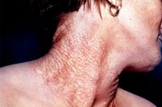
Clinically manifested by flat, yellowish, grouped papules measuring 1-3 mm, often located along the skin lines on the lateral surfaces of the neck, back of the head, in the armpits and groin areas, on the abdomen, in the popliteal fossa, on the elbows. The surface of the papules is smooth, the skin in the areas of the rash is flabby, often forming folds, which makes it indistinguishable from flabby skin. Mucous membranes may be affected. Changes in the eyes consist of slowly progressing dystrophic changes in the fundus, resulting from the divergence and rupture of the basal plate (Bruch's elastic membrane), localized between the vascular membrane and the retina. This leads to the formation of so-called angioid streaks. They are revealed during examination of the fundus as jagged lines or stripes with pigmentation. Angioid streaks are not specific for pseudoxanthoma elasticum, they are also found in Chernogubov-Eders-Danlos syndrome, Paget's disease, Marfan syndrome and sickle cell anemia. They may be the only sign of pseudoxanthoma elasticum for many years. Angioid streaks are often combined with hemorrhages under the retina and choroid, as well as with retinal detachment. In 50% of patients, punctate changes are observed leading to a significant decrease in vision. Cardiovascular lesions are characterized by hypertension and coronary insufficiency, early atherosclerosis, and a tendency to hemorrhage. In the same family, siblings may have mono-, di- and tri-symptom forms of the disease. The severity of skin and eye symptoms varies significantly.
What do need to examine?
How to examine?
Treatment of an elastic pseudoxanthoma
Currently, there is no effective specific treatment for pseudoxanthoma elasticum. In stage I of the ophthalmologic lesion, observation is prescribed, it is recommended to avoid the slightest eye injury and wear protective glasses during work and sports. Treatment of stage II presents significant difficulties. There are studies on the use of laser coagulation of angioid streaks tending to the macular zone. Intravitreal injections of monoclonal antibodies that block angiogenesis (for example, bevacizumab) are promising for the treatment of angioid streaks of the retina. However, reliable data on the effectiveness of this treatment method have not been obtained. In stage III, treatment is ineffective. The goal of therapy is to prevent complications.