Medical expert of the article

New publications
Ectomesodermal focal dysplasia
Last reviewed: 07.07.2025

All iLive content is medically reviewed or fact checked to ensure as much factual accuracy as possible.
We have strict sourcing guidelines and only link to reputable media sites, academic research institutions and, whenever possible, medically peer reviewed studies. Note that the numbers in parentheses ([1], [2], etc.) are clickable links to these studies.
If you feel that any of our content is inaccurate, out-of-date, or otherwise questionable, please select it and press Ctrl + Enter.
Focal ectomesodermal dysplasia (syn.: Goltz syndrome, Goltz-Gorlin syndrome, focal dermal hypoplasia, mesoectodermal dysplasia syndrome) is a rare disease, probably genetically heterogeneous, but in most cases inherited in an X-linked dominant manner with a lethal outcome in male fetuses, gene locus Xp22.31. It occurs almost exclusively in women.
 [
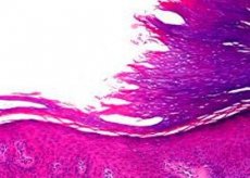
[ Pathomorphology of focal ectomesodermal dysplasia
In atrophic areas of the skin, thinning of the dermis is observed with a normal epidermis structure. The subcutaneous fat layer is located close to the epidermis, sometimes at the level of the superficial vascular plexus. The number of vessels in the papillary layer is increased, sparseness of fibrous structures is noted. Collagen fibers are thinned, located in the form of individual threads. There are few skin appendages, but their structure is within normal limits. This picture resembles the superficial lipomatous nevus of the skin of Hoffman-Zurkale. In areas of hypoplasia with fatty protrusions, a similar, but more pronounced picture is revealed. In the area of papillomatous growths of the mucous membranes, the histological picture corresponds to a papilloma with increased vascularization of the stroma. Electron microscopic examination of atrophic skin areas revealed fibroblasts with long cytoplasmic outgrowths and poorly developed endoplasmic reticulum, around which are located thin fibrils without transverse striation, possibly complexes of tropocollagen and procollagen. Mature collagen fibers do not differ in structure from normal ones. In the subcutaneous base, fat cells with many fat vacuoles predominate - young forms indicating proliferation of fat cells.
The histogenesis of skin changes in focal ectomesodermal dysplasia is associated with a disruption in the kinetics of fibroblast growth during their normal synthetic activity, resulting in a paucity of connective tissue elements of the dermis. At the same time, there is a proliferation of subcutaneous tissue cells that replace the defective dermis.
Symptoms
Clinically, it manifests itself as atrophopoikilodermic changes in the skin with herniated protrusions of the subcutaneous tissue, anomalies of the skeleton, teeth and eyes, appearing from birth. Papillomas on the mucous membranes of the lips, vagina, anus, lesions of the skin appendages in the form of nail dystrophy, hair growth disorders with nesting alopecia and sweating are often observed.
Mental retardation with epileptiform seizures is noted.
What do need to examine?
How to examine?