Medical expert of the article

New publications
ECG with exercise: how to do, normal values, interpretation
Last reviewed: 04.07.2025

All iLive content is medically reviewed or fact checked to ensure as much factual accuracy as possible.
We have strict sourcing guidelines and only link to reputable media sites, academic research institutions and, whenever possible, medically peer reviewed studies. Note that the numbers in parentheses ([1], [2], etc.) are clickable links to these studies.
If you feel that any of our content is inaccurate, out-of-date, or otherwise questionable, please select it and press Ctrl + Enter.
The study of the electrical activity of cardiac muscle cells – exercise ECG – evaluates the ability of the myocardium to respond to physical exercise in a controlled clinical environment. With exercise ECG, cardiologists are able to obtain key parameters of the heart’s functioning under conditions close to natural, since the patient’s body is in motion.
The exercise stress test compares the coronary circulation of the same patient at rest and under physical exertion, showing the frequency, regularity and duration of heart contractions and the ability of the cardiovascular system to withstand stress and provide blood flow to the myocardium.
And the results of this study can reflect both the general physical condition of a person and indicate cardiovascular pathologies, primarily coronary heart disease.
 [
[ Indications for the procedure
Healthy people undergo stress ECG during periodic examinations of professional athletes, civil and military aviation flight personnel. Such electrocardiography is performed on candidates for contract service in the army, special forces units of law enforcement agencies and rescue services.
An ECG with physical activity for children is required either to assess the ability to engage in a particular sport, or to clarify the reasons for a child or teenager's complaints of rapid heartbeat and pain in the heart area.
Indications for performing a stress ECG for diagnostic purposes include:
- ischemic heart disease, and if present, monitoring of the myocardial condition;
- monitoring the state of cardiac activity in patients who have had a heart attack or coronary artery bypass grafting;
- heart valve defects (chronic aortic regurgitation );
- sinus arrhythmia;
- coronary artery stenosis;
- disturbances of atrioventricular conduction (atrioventricular heart block), etc.
The corresponding ECG parameters with exercise stress – taking into account the results of other examinations – either serve as confirmation of the diagnosis or may be an objective basis for its exclusion.
In addition, this study of the work of the heart muscle helps to evaluate the effectiveness of a specific program for the treatment of cardiovascular diseases, as well as to establish the limits of acceptable, safe loads for the heart before starting rehabilitation after myocardial infarction or cardiac surgery (bypass surgery, angioplasty).
If necessary, the doctor you contact will give you a referral for examination and tell you where to do an ECG with physical activity (in the same medical institution or any other).
Preparation
Preparation for this study means that the patient should not drink caffeinated drinks, alcohol, or chocolate, or smoke for 24 hours before the test. The last meal should be three to four hours before the procedure. Physical activity should also be avoided for at least two days.
In addition, when prescribing an ECG stress test with physical exertion, the doctor warns male patients to stop taking any medications to improve erection (Viagra, Cialis, Levitra, etc.) three days before.
Patients should also inform their doctor about all medications they are taking, in particular cardiotonic and antiarrhythmic drugs, to avoid distorted ECG results.
Technique ECG with exercise: how to do, normal values, interpretation
The technique for performing a stress electrocardiological test depends on the type of physical activity:
- regular squats (at least 20 in 45-60 seconds),
- step platforms (lowering and rising with both legs with the same intensity),
- on a treadmill (running at a moderate pace for 20-25 seconds),
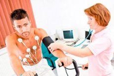
- on a bicycle ergometer (a computerized exercise bike, the pedals of which must be rotated at a certain number of revolutions for three minutes). In addition to heart function readings, changes in blood pressure are simultaneously recorded during exercise using a bicycle ergometer (for which a blood pressure cuff is placed on the arm).
How is an ECG performed with exercise? Regardless of the technical component of the study, the procedure begins with the installation of 6-9 electrodes on the chest (in clearly defined places - at the left and right edges of the sternum, at the left armpit, etc.). Through these electrodes, the electrocardiograph will take readings (potential difference in leads) and record them on the electrocardiogram. Readings are taken twice - an ECG at rest and with exercise: a regular ECG (in the supine position) is needed to obtain neutral indicators with which the parameters of electrical activity of myocardial cells during physical exercise will be compared.
The health worker monitors the patient's condition both during and after testing until the heart rhythm returns to normal.
Contraindications to the procedure
Among the contraindications to electrocardiography with physical activity, experts note:
- recent acute myocardial infarction;
- severe heart defects;
- decompensated or inadequately controlled congestive heart failure;
- acute coronary syndrome;
- severe unstable angina;
- severe cardiac arrhythmia, such as ventricular tachycardia;
- dysfunction of one or more heart valves;
- severe aortic stenosis, cardiac aneurysm with aortic dissection;
- acute pulmonary embolism;
- hypertrophic cardiomyopathy;
- any inflammatory diseases of the heart ( pericarditis, myocarditis, endocarditis );
- acute cerebrovascular accident;
- hypertension stage III;
- inflammation of the venous walls with the formation of blood clots;
- presence of a pacemaker.
Normal performance
If after 20-30 squats (their specific number depends on the age of the patients), performed for one minute, the heart rate (the norm at rest is 60-90 beats/min) increases within 20%, then this is the norm of the ECG with load. After all, an increase in pulse rate and an increase in blood pressure is a healthy reaction of the cardiovascular system to physical exertion and means that the heart copes with pumping blood. The definition of the rhythm as sinus also means the norm.
An increase in heart rate by 30-50% indicates a decreased cardiac endurance, and, therefore, problems with its functioning. Experts note that when interpreting electrocardiography results, the conclusion about the presence of ischemic heart disease (in particular, subendocardial) is determined by such ECG indicators with a load as horizontal depression of the ST segment (in leads V4, V5 and V6); coronary insufficiency is indicated by ventricular arrhythmia against the background of the same depression of the ST segment, and unstable angina is indicated by changes in T-waves and the position of the T wave on the isoelectric line of the ECG.
Patients should understand that the description of the ECG conclusion with exercise (as well as a regular ECG) is information for cardiologists, which provides grounds for conclusions about the condition of the heart and diagnosis. Only specialists in the field of electrocardiography are engaged in its decoding, who are not obliged to explain to patients what the terms indicated in the ECG conclusion mean (P and T waves, RR, ST, PQ intervals, etc.). Or that chest leads are electrocardiogram curves recorded from electrodes attached to the chest, and the QRS complex is called the excitation period of the ventricles of the heart pumping blood...
However, the physician should explain to the patient the main parameters of the exercise ECG. ST segment changes, ventricular arrhythmia, and T-wave abnormalities do not necessarily represent a positive result. Moreover, if the exercise ECG does not reach 85% of the maximum heart rate, then a negative result has no diagnostic value. However, with a positive result, the probability of myocardial ischemia is almost 98%.
Complications after the procedure
During the ECG stress test with physical exertion, the patient may experience fatigue, dizziness, difficulty breathing, rapid heartbeat, chest discomfort, leg pain. The doctor should be informed about this to prevent possible complications after the procedure, when vegetative symptoms increase (impaired coordination of movements, intention tremor, leg cramps); signs of impaired pulmonary ventilation and perfusion occur ( shortness of breath, wheezing, pale skin, cyanosis); persistent ventricular tachycardia is noted; chest pain increases.
In the presence of myocardial ischemia, a hypertensive response develops with an increase in systolic blood pressure above 250 mm Hg in response to increased physical activity.
Cardiovascular problems cause consequences after the procedure in the form of: atrial fibrillation, ventricular tachycardia and ventricular fibrillation, conduction disturbances, acute heart failure and myocardial infarction; bronchospasm (in bronchial asthma due to physical effort); fainting or stroke.