Medical expert of the article

New publications
Duplex scanning of lower limb veins
Last reviewed: 29.06.2025

All iLive content is medically reviewed or fact checked to ensure as much factual accuracy as possible.
We have strict sourcing guidelines and only link to reputable media sites, academic research institutions and, whenever possible, medically peer reviewed studies. Note that the numbers in parentheses ([1], [2], etc.) are clickable links to these studies.
If you feel that any of our content is inaccurate, out-of-date, or otherwise questionable, please select it and press Ctrl + Enter.
Ultrasound duplex scanning, or ultrasound duplex scanning of the veins of the lower extremities, gives the doctor the opportunity to trace the basic values of hemodynamics - in particular, the direction and speed of blood flow, the degree of vascular filling, as well as to give an objective assessment of the state of the venous network and surrounding structures. This procedure is completely safe and virtually devoid of contraindications. The study is performed during pregnancy and in early childhood, without restrictions.
Indications
The main indication for the appointment of duplex scanning of the veins of the lower extremities is swelling of the legs, regular muscle cramps, painful sensations, darkening or lightening of the skin on the legs, the appearance of spots, structural changes in the skin, sharp periodic pain, inflammatory processes along the course of the venous channel.
A duplex scan can detect:
- Deep vein varicose veins;
- Insufficient function of the venous vessels of the lower extremities;
- Clotting;
- Inflammatory processes in the veins;
- Venous fibrosis;
- Blood flow disorder due to external compression of blood vessels;
- Disturbed combination of venous and arterial network;
- Genetic and congenital developmental abnormalities.
Preparation
Duplex scanning of the lower extremity veins does not require any major preparation.
In order for the results of the examination to be more correct, the day before the examination should be avoided:
- Heavy physical exertion;
- Smoking and drinking alcohol;
- The use of stimulant drinks, coffee.
The possibility of taking certain medications before diagnosis is determined by the attending physician.
Technique
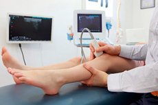
How is a duplex scan of the lower limb veins performed? This procedure is not complicated and is suitable for people of different ages and health conditions. Both children and pregnant women undergo the scan without any unpleasant sensations or consequences.
Before the procedure it is not necessary to remove all clothing, but it is necessary to expose the lower extremities (remove pants, tights, socks).
The specialist will suggest which position the patient should take, and then apply a special contact gel to the skin or directly to the ultrasound transducer.
During the scanning process, the doctor guides the transducer over the desired area of the limb. At the same time, you can see a picture of the area being scanned on the monitor screen.
Sometimes during one procedure the patient has to change the position of the body several times: for example, first the doctor will ask to stand upright, then - lie on the stomach (for better visualization of the hamstring or tibial arteries), bend or straighten the leg.
There are practically no contraindications to ultrasound duplex scanning. Possible exception - dermatologic diseases and injuries in the area of the proposed study.
What does a duplex scan of the lower extremity veins show?
The method of duplex scanning of lower limb veins helps to assess the degree of patency of deep and superficial vessels, inferior vena cava and external iliac veins. During the procedure, it is possible to distinguish thrombotic accumulations that are not visible in the conventional B-mode.
Color duplex scanning of lower limb veins is a rather lengthy procedure that lasts about 40-60 minutes. The specialist determines the state of the venous lumen, valve system, and then performs a number of functional tests that provide information about the direction of blood flow, the presence of pathological opposite blood discharge, the state of connections and joints.
In the process of ultrasound duplex diagnostics, the quality of blood circulation in the superficial veins of the thigh is determined, a test for the functionality of the terminal and preterminal valve of the great saphenous vein is performed. Evaluate the state of the superficial, deep, common veins of the thigh. Valsalva test is performed, which determines the functionality of venous valves of the inguinal-femoral region. The essence of this test is as follows: the patient deeply inhales, and then holds his breath, while trying to maximize pushing. If the valve system is not working properly, the doctor will fix the opposite blood flow in the projection area of the ultrasound sensor.
The same principle is used for ultrasound duplex examination of the common, deep, superficial femoral and hamstring veins.
Duplex scanning of the lower leg vessels is performed standing up, which is associated with the greatest tension of the venous walls and the load on the valves in the upright position.
In addition, the functionality of the connecting veins is assessed in the ankle and thigh joints.
The use of color and spectral scans provides essential information about the state of the venous network and the extent of the pathological response.
If the doctor suspects venous thrombosis of the lower extremities, he carefully examines the deep veins, describes the location and structure of the blood clots. Correct interpretation of the results helps to further choose the right therapeutic tactics. Early diagnosis can avoid surgical intervention, or extremely delay it.
Decoding of duplex scanning of lower limb veins
The venous system of the lower extremities is divided into two categories: superficial and deep veins. Both vessels are equipped with valves that provide one direction of blood flow: from the periphery to the center, or from bottom to top.
The valves are bicuspid, localized up to 10 cm apart. The deep venous network provides an outflow of 85-90% of blood, and the superficial one - 10-15% of blood. All veins of the lower extremities have several layers: endothelial, middle (muscular) and connective tissue (collagen-elastin). The outer layer also contains collagen, which provides strength to the wall. The connection between the superficial and deep veins is provided by connective small vessels with a diameter size of about 2 mm and a length of no more than 150 mm. In each of them there are 1-2 valves. The direction of blood flow in the connections is from the superficial network to the deep network.
A specialist can identify such causes of venous deficiency:
- Impairment of contractile cardiac function and the associated reduction in the influence of the right atrial pumping factor;
- Loss of venous patency;
- Failure of the mechanism of venous outflow in the standing position.
Among the underlying causes of poor venous function are varicose veins (dilated veins) and post-thrombotic syndrome.
Normal when the venous contour is smooth, with a gradual increase in diameter in the proximal direction. Uniformly dilated areas are detected in areas with valves. The vein wall itself has a dense structure, so it is tinged with a white color. The vascular lumen, inside which there is blood, is hypoechogenic, so the image is darkened.
The diameter of the vein is not constant and may vary depending on posture, respiratory phase, individual characteristics, etc.
Duplex scanning of the veins of the lower extremities demonstrates the condition of all veins on all sides of the leg, allows you to identify the relationship of blood flow and respiratory movements, the possibility of compression of the connecting vessels, as well as detect stenoses, thrombi, reflux pathologies, non-functioning veins.