Medical expert of the article

New publications
Duodenal probing of the gallbladder
Last reviewed: 06.07.2025

All iLive content is medically reviewed or fact checked to ensure as much factual accuracy as possible.
We have strict sourcing guidelines and only link to reputable media sites, academic research institutions and, whenever possible, medically peer reviewed studies. Note that the numbers in parentheses ([1], [2], etc.) are clickable links to these studies.
If you feel that any of our content is inaccurate, out-of-date, or otherwise questionable, please select it and press Ctrl + Enter.

 [
[ Indications for the procedure
This study is used in the diagnosis of diseases of the gallbladder and bile ducts, duodenum. However, at present, due to the widespread use of endoscopy and ultrasound, this method is used less often. The contents of the duodenum are a mixture of bile, pancreatic and duodenal secretions with a small amount of gastric juice.
Multi-stage fractional duodenal sounding allows obtaining bile from the common bile duct, gall bladder and intrahepatic bile ducts with subsequent biochemical and microscopic examination. In addition, this method provides an idea of the functional state of the gall bladder and bile ducts.
Preparation
Before inserting the tube, a pharyngeal swab should be taken for bacteriological examination, then the patient should rinse the oral cavity with a disinfectant solution to reduce the possibility of introducing microflora from the oral cavity into portions of bile. The duodenal tube is inserted into the duodenum in the morning on an empty stomach. It is more preferable to use a two-channel tube of N. A. Skuya for separate extraction of gastric and duodenal contents. One channel of the tube is located in the stomach, the other - in the duodenum. Gastric juice should be continuously extracted with a syringe or vacuum unit, since when hydrochloric acid of gastric juice enters the duodenum, bile becomes cloudy. In addition, hydrochloric acid stimulates pancreatic secretion and bile excretion due to the release of secretin and cholecystokinin-pancreozymin hormones.
If a two-channel probe is not available, a single-channel duodenal probe should be used.
The device for carrying out the procedure
The examination is best performed with a two-channel probe with a metal olive with holes at the end. There are 3 marks on the probe: at a distance of 45 cm (the distance from the incisors to the subcardial part of the stomach), 80 cm (the distance to the large duodenal papilla).
Fractional duodenal intubation (FDS) has the following advantages over conventional duodenal intubation:
- allows you to get a clearer idea of the functional state of the gallbladder and bile ducts;
- allows to diagnose the type of gallbladder dyskinesia.
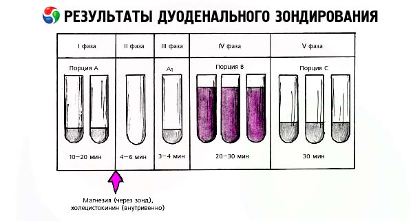
Technique duodenal probing
Collection of bile from duodenal contents is carried out in numbered test tubes every 5 minutes.

There are 5 phases of fractional duodenal sounding.
- 1 - choledochochus phase - begins after the probe olive is located in the duodenum (angle of the descending and lower horizontal part). During this period, the sphincter of Oddi is in a relaxed state and a portion of transparent light yellow bile is released from the common bile duct (d. choledochus) as a result of irritation of the duodenum by the probe olive.
The time during which bile is secreted and its volume are taken into account.
Phase 1 reflects the basal secretion of bile (outside digestion) and the partially functional state of the sphincter of Oddi.
Normally, 15-20 ml of bile is secreted within 10-15 minutes (according to some data - within 20-40 minutes).
After the end of the secretion of bile into the duodenum, a warm 33% magnesium sulfate solution heated to 37°C is slowly introduced through the duodenal tube over 5-7 minutes - 30 ml or 5% - 50 ml.
In response to the introduction of the stimulus, the sphincter of Oddi reflexively closes and remains closed throughout the second phase of probing.
- Phase 2 - closed sphincter of Oddi (the latent period phase of bile secretion) - reflects the time from the introduction of the cholecystokinetic solution to the appearance of bile-stained secretion. At this time, bile is not secreted. This phase characterizes the cholestatic pressure in the biliary tract, the readiness of the gallbladder to empty and its tone.
Normally, the phase of the closed sphincter of Oddi lasts 3-6 minutes.
If bile appears before 3 minutes, this indicates hypotension of the sphincter of Oddi. An increase in the time of the closed sphincter of Oddi for more than 6 minutes indicates an increase in its tone or a mechanical obstruction of the outflow of bile. To resolve the issue of the nature of the changes, 10 ml of warm (heated to 37 ° C) 1% novocaine solution can be administered through a tube. The appearance of light yellow bile after this indicates a spasm of the sphincter of Oddi (novocaine relieves the spasm). If bile is not released within 15 minutes after the introduction of novocaine, the patient can be given 1/2 a nitroglycerin tablet under the tongue and, if there is no effect, a cholekinetic agent (20 ml of vegetable oil or 50 ml of a 40% glucose solution, xylitol) can be reintroduced through a tube into the duodenum. If bile does not appear after this, the position of the probe in the duodenum should be checked radiologically, and if the probe is positioned correctly, stenosis in the area of the d. choledochus can be assumed.
- Phase 3 - A-bile (cystic duct phase) - begins with the opening of the sphincter of Oddi and the appearance of light bile A until the release of dark concentrated bile from the gallbladder.
Normally, this period lasts 3-6 minutes, during which 3-5 ml of light bile is released from the cystic and common bile ducts.
This phase reflects the state of these ducts. An increase in the time of phase 3 over 7 minutes indicates an increase in the tone of the Lutkens sphincter (it is located at the transition of the neck of the gallbladder to the cystic duct) or hypotension of the gallbladder.
Gallbladder hypotension can only be discussed after comparing data from stages III and IV.
Bile of phases 1, 2 and 3 constitutes the classic portion A of conventional (non-fractional) duodenal sounding.
- Phase 4 - gallbladder (cystic bile, B-bile phase) - characterizes the relaxation of the Lutkens sphincter and emptying of the gallbladder.
Phase 4 begins with the opening of the Lutkens sphincter and the appearance of dark olive concentrated bile and ends when the secretion of this bile stops.
The secretion of gallbladder bile is initially very intense (4 ml per minute), then gradually decreases.
Normally, the gallbladder takes 20-30 minutes to empty, during which time an average of 30-60 ml of dark olive gallbladder bile is released (with chromatic probing, the bile is colored blue-green).
Intermittent secretion of gallbladder bile indicates dyssynergism of the sphincters of Lutkens and Oddi. An increase in the time of gallbladder bile secretion (more than 30 minutes) and an increase in the amount of more than 60-85 ml indicate gallbladder hypotension. If the duration of phase 4 is less than 20 minutes and less than 30 ml of bile is secreted, this indicates hypertonic dyskinesia of the gallbladder.
- Phase 5 - the phase of hepatic bile-C - occurs after the end of the secretion of B-bile. Phase 5 begins with the secretion of golden bile (hepatic). This phase characterizes the exocrine function of the liver. During the first 15 minutes, hepatic bile is secreted intensively (1 ml or more per 1 minute), then its secretion becomes monotonous (0.5-1 ml per 1 minute). Significant secretion of hepatic bile in phase 5, especially in the first 5-10 minutes (>7.5 ml/5 min) indicates the activity of the sphincter of Mirizzi, which is located in the distal part of the hepatic duct and prevents the retrograde movement of bile during contraction of the gallbladder.
Bile - It is advisable to collect it for 1 hour or more, studying the dynamics of its secretion, and try to obtain residual gallbladder bile without re-introducing a gallbladder irritant.
Repeated contraction of the gallbladder normally occurs 2-3 hours after the introduction of the irritant. Unfortunately, in practice, duodenal intubation is completed 10-15 minutes after the appearance of hepatic bile.
- Many suggest distinguishing phase 6 - the phase of residual gallbladder bile. As indicated above, 2-3 hours after the introduction of the irritant, the gallbladder contracts again.
Normally, the duration of phase 6 is 5-12 minutes, during which time 10-15 ml of dark olive gallbladder bile is secreted.
Some researchers suggest not to wait 2-3 hours, but to introduce an irritant soon after obtaining liver bile (after 15-20 minutes) to be sure of complete emptying of the gallbladder. Obtaining additional amounts of gallbladder (residual) bile during this period of time indicates incomplete emptying of the gallbladder during its first contraction and, consequently, its hypotension.
Normal performance
For a more detailed study of the function of the sphincter apparatus of the biliary tract, it is advisable to study bile secretion graphically, with the volume of bile obtained expressed in ml, and the time of bile secretion in min.
It is proposed to determine a number of indicators of bile secretion:
- the rate of bile secretion from the bladder (reflects the efficiency of bile release by the bladder) is calculated using the formula:
H=Y/T, where H is the rate of bile secretion from the gallbladder; V is the volume of gallbladder bile (B-portion) in ml; T is the time of bile secretion in min. Normally, the rate of bile secretion is about 2.5 ml/min;
- The evacuation index is an indicator of the motor function of the gallbladder and is determined by the formula:
IE = H/Vостат*100%. IE is the evacuation index; H is the rate of bile secretion from the gallbladder; Vостат is the residual volume of gallbladder bile in ml. Normally, the evacuation index is about 30%;
- the effective release of bile by the liver is determined by the formula:
EVL = V portion of bile C in 1 hour in ml / 60 min, where EVL is the effective release of hepatic bile. Normally, EVL is about 1-1.5 ml/min;
- The secretory pressure index of the liver is calculated using the formula:
The index of secretory pressure of the liver = EEJ/H * 100%, where EEJ is the effective release of hepatic bile; H is the rate of secretion of hepatic bile from the bladder (effective release of bile by the bladder). Normally, the index of secretory pressure of the liver is approximately 59-60%.
Fractional duodenal sounding can be made chromatic. For this purpose, the day before duodenal sounding at 2100, 2 hours after the last meal, the patient takes 0.2 g of methylene blue in a gelatin capsule orally. The next morning at 9:00 (i.e. 12 hours after taking the dye), fractional sounding is performed. Methylene blue, having been absorbed in the intestine, enters the liver with the bloodstream and is reduced in it, turning into a colorless leuco compound. Then, having entered the gallbladder, the discolored methylene blue oxidizes, turns into a chromogen and colors the gallbladder bile blue-green. This allows one to confidently distinguish gallbladder bile from other phases of bile that retain their normal color.
The bile obtained during duodenal intubation is examined biochemically, microscopically, and bacterioscopically; its physical properties and the sensitivity of the flora to antibiotics are determined.
Bile should be examined immediately after its collection, since the bile acids it contains quickly destroy formed elements. Bile should be delivered to the laboratory warm (test tubes with bile are placed in a jar with warm water), so that lamblia can be more easily detected under microscopy (in cold bile they lose their motor activity).
Changes in duodenal sounding parameters (portion "B"), characteristic of chronic cholecystitis
- The presence of a large number of leukocytes, especially the detection of their clusters. The question of the diagnostic value of detecting leukocytes in bile as a sign of an inflammatory process has not been finally resolved. Leukocytes can enter any portion of the duodenal contents from the mucous membrane of the oral cavity, stomach, and duodenum. Leukocytoids, cells of the cylindrical epithelium of the duodenum that have transformed into large round cells resembling leukocytes under the influence of magnesium sulfate, are often mistaken for leukocytes. In addition, it should be taken into account that leukocytes are quickly digested by bile, which, of course, reduces their diagnostic value.
In this regard, it is currently believed that the detection of leukocytes in portion B is a sign of an inflammatory process only if the following conditions are present:
- if the number of leukocytes is really high. To identify leukocytes, one should use Romanovsky-Giemsa staining, and also conduct a cytochemical study of the peroxidase content in the cells. Leukocytes give a positive reaction to myeloperoxidase, leukocytoids do not;
- if accumulations of leukocytes and columnar epithelial cells are found in mucus flakes (mucus protects leukocytes from the digestive action of bile);
- if the detection of leukocytes in bile is accompanied by other clinical and laboratory signs of chronic cholecystitis.
The detection of leukocytoids is not given diagnostic value. To detect leukocytes and other cells in bile, at least 15-20 preparations should be examined under a microscope.
- Visual examination of bile reveals its pronounced turbidity, flakes and mucus. In a healthy person, all portions of bile are transparent and do not contain pathological impurities.
- Detection of a large number of columnar epithelial cells in bile. It is known that three types of columnar epithelium can be detected in bile: small epithelium of the intrahepatic bile ducts - in cholangitis (in portion "C"); elongated epithelium of the common bile duct when it is inflamed (portion "A"); broad epithelium of the gallbladder in cholecystitis.
Chronic cholecystitis is characterized by the detection of a large number of columnar epithelial cells (mostly wide) in the gallbladder bile. The columnar epithelial cells are found not only as individual cells, but also in clusters (layers) of 25-35 cells.
- Decrease in the pH of gallbladder bile. Gallbladder bile normally has a pH of 6.5-7.5. In inflammatory diseases of the biliary system, the reaction becomes acidic. According to researchers, in the case of exacerbation of chronic cholecystitis, the pH of gallbladder bile can be 4.0-5.5.
- The appearance of cholesterol and calcium bilirubinate crystals. Chronic cholecystitis is characterized by the appearance of cholesterol and calcium bilirubinate crystals. The detection of a large number of them indicates destabilization of the colloidal structure of bile (dyscrinia). When conglomerates of these crystals and mucus appear, one can talk about the lithogenic properties of bile, the formation of microliths and a peculiar transformation of non-calculous cholecystitis into calculous. Together with microliths, "sand" is often found - small grains of various sizes and colors (colorless, refracting light, brown), recognizable only under a microscope, which are located in mucus flakes.
- Decreased relative density of gallbladder bile. Normally, the relative density of gallbladder bile is 0.016-1.035 kg/l. In severe exacerbation of chronic cholecystitis, a decrease in the relative density of gallbladder bile is observed due to its dilution by inflammatory exudate.
- Changes in the biochemical composition of bile. Bile is a complex colloidal solution containing cholesterol, bilirubin, phospholipids, bile acids and their salts, minerals, proteins, mucoid substances, and enzymes.
During an exacerbation of chronic cholecystitis, the biochemical composition of bile changes:
- the amount of mucin substances that react with the DPA reagent increases, which significantly increases the activity of the DPA reaction;
- the content of glycoproteins (hexosamines, sialic acids, fucoses) in bile increases by 2-3 times;
- the content of bile acids decreases;
- the cholate-cholesterol ratio (the ratio of the content of bile acids in bile to the level of cholesterol in it) decreases;
- the content of lipoprotein (lipid) complex decreases.
Lipoprotein macromolecular complex is a complex compound formed in the liver, which includes the main components of bile: bile acids, phospholipids, cholesterol, bilirubin, protein, grouped around lipoprotein cores to form a macromolecular complex. The lipoprotein complex ensures colloidal stability of bile and its flow from the liver to the intestine. Bile phospholipids form micelles with cholesterol, and bile acids stabilize them and convert cholesterol into a soluble form;
- the content of fibrinogen and its metabolic products in the gallbladder bile increases sharply;
- proteinocholia is observed - increased secretion of serum proteins (mainly albumins) into bile with a simultaneous decrease in the content of secretory immunoglobulin A.
- Increased content of lipid peroxides in gallbladder bile.
The increase in the amount of lipid peroxides in bile is a consequence of the sharp activation of free radical oxidation of lipids. The level of lipid peroxides clearly correlates with the severity of the inflammatory process in the gallbladder.
- Bacteriological examination of bile. The purpose of bacteriological examination of bile is to detect bacterial flora and determine its sensitivity to antibacterial agents. The study has diagnostic value if the number of bacteria exceeds 100,000 in 1 ml of bile.