Medical expert of the article

New publications
Gallbladder empyema
Last reviewed: 04.07.2025

All iLive content is medically reviewed or fact checked to ensure as much factual accuracy as possible.
We have strict sourcing guidelines and only link to reputable media sites, academic research institutions and, whenever possible, medically peer reviewed studies. Note that the numbers in parentheses ([1], [2], etc.) are clickable links to these studies.
If you feel that any of our content is inaccurate, out-of-date, or otherwise questionable, please select it and press Ctrl + Enter.
A condition in which a large amount of purulent discharge accumulates in the gallbladder without the possibility of its release is called empyema of the gallbladder. Bacterial infection and blockage of the cystic duct play a role in the development of this pathology. The disease reveals itself with severe pain, high temperature and increasing signs of intoxication.
Empyema of the gallbladder is most often one of the unfavorable consequences of the acute inflammatory process - cholecystitis. The main difference between empyema and purulent cholecystitis is the disruption of the outflow of bile caused by obstructive blockage of the duct. The complication occurs in approximately 10% of patients with acute cholecystitis. [ 1 ]
Epidemiology
The actual extent of gallbladder empyema is difficult to trace. However, according to information obtained in the course of several studies, the occurrence of this complication is noted in approximately 5-15% of patients with cholecystitis. The most common cause of the development of pathology is unresolved acute calculous cholecystitis.
Empyema of the gallbladder is one of the serious complications of acute cholecystitis. Other possible complications include gangrenous cholecystitis, dropsy, and gallbladder perforation. Perforation occurs in approximately 6-12% of cases of acute cholecystitis, with a mortality rate of 20-24% (while with gangrenous cholecystitis it is 20%).
Empyema of the gallbladder most often affects people over 50 years of age, but the disease also occurs at a younger age. Elderly and senile patients make up about 45-50% of the total number of patients. Men and women get sick with approximately the same frequency. [ 2 ]
Causes gallbladder empyema
Empyema of the gallbladder is not a primary disease: it is always secondary and occurs as a complication of some other, initial pathology. The main causes of empyema are:
- acute inflammatory processes in the biliary system (cholecystitis with or without the formation of stones), creating obstacles to the excretion of bile, leading to stagnation and increased growth of bacterial flora; [ 3 ]
- tumor processes that compress the bile duct, preventing the excretion of bile.
The development of empyema is most often provoked by the following types of microorganisms:
- Escherichia coli;
- Klebsiella pneumoniae;
- Streptococcus faecalis;
- bacteroides;
- Clostridium spices.
Empyema of the gallbladder develops more rapidly in patients suffering from obesity, diabetes mellitus, immunodeficiency states and hemoglobinopathies, as well as biliary carcinoma.
The role of the allergic predisposition of the organism is also taken into account in pathogenesis. Local allergic effects on the biliary tract of bacterial toxins, medications, and chemicals aggravate the already impaired function of the organ. Parasitic invasions (in particular, opisthorchiasis) can cause the development of cholecystitis, increase the virulence of bacteria, contribute to allergic manifestations, motility disorders, and the development of congestion. [ 4 ]
Risk factors
Empyema of the gallbladder occurs as a result of immediate causes of acute inflammatory process – cholecystitis. However, other disorders of body functions that can become a catalyst – a trigger for the development of inflammation should not be ignored. [ 5 ]
Such risk factors include the following:
- frequent or chronic otolaryngological and respiratory diseases, including sinusitis, bronchitis, sinusitis, pneumonia, etc.;
- chronic or acute inflammatory processes of the digestive system (enterocolitis, appendicitis, intestinal microflora disorders, etc.);
- parasitic diseases, helminthiasis;
- infections of the genitourinary system (pyelonephritis, salpingoophoritis, cystitis, prostatitis, etc.);
- biliary dyskinesia, gallbladder tone disorders, cholelithiasis;
- poor nutrition (especially regular overeating or fasting, as well as abuse of spicy, fatty and fried foods);
- autoimmune diseases;
- tumors;
- arterial hypertension, diabetes mellitus and other pathologies that can indirectly disrupt the blood supply to the hepatobiliary system;
- hormonal changes, including during pregnancy;
- obesity, metabolic disorders;
- alcohol and tobacco abuse;
- severe or frequent allergic reactions;
- predominantly sedentary lifestyle;
- genetic predisposition.
According to statistics, a considerable number of cases of acute cholecystitis, which can provoke the development of empyema of the gallbladder, occur against the background of the presence of gallstones. Gallstone disease is one of the leading risk factors for the development of the disease.
Another factor that is rarely mentioned by specialists is a woman’s prolonged and difficult labor, which can cause damage to the gallbladder and significantly increase the likelihood of an inflammatory process developing already in the early postpartum stage.
Bladder injuries can occur not only during childbirth, but also in everyday life. In this case, almost any mechanical damage to the abdominal cavity, and especially to the right hypochondrium, becomes dangerous.
Uncompensated diabetes mellitus increases the risk of inflammation and damage to the biliary system.
Common prerequisites for gallbladder dysfunction may be nutritional disorders, failure to adhere to a dietary regimen, overeating or eating too infrequently, excessive consumption of fried and fatty foods, alcohol, as well as psychoemotional, allergic and other negative phenomena, including infectious pathologies.
During the examination of practically healthy volunteers, specialists determined that the fasting gallbladder volume directly correlates with a person's weight. But disorders of the motor function of the biliary system were found only in people with excess weight and increased gallbladder volume on an empty stomach, which indicates the involvement of obesity in the development of disorders of the biliary system. Some scientists also associate the development of pathology with a deficiency of vitamin D 2 and metabolic disorders.
Pathogenesis
Empyema of the gallbladder occurs against the background of blocked bile flow and the addition of an infectious component. The blockage may be caused by the wedging of stones into the bladder neck, blockage of the duct by a bile clot, or compression by a nearby tumor process. Acute cholecystitis becomes the trigger factor. [ 6 ]
Gallbladder inflammation develops when an infection enters – through the bloodstream, lymph flow or from the intestinal cavity. If the motility of the bile duct is impaired, microorganisms can penetrate the biliary system from the intestine.
The presence of stones, kinks or narrowing of the duct leads to stagnation of bile in the organ. In approximately 90% of cases, acute cholecystitis occurs due to cholelithiasis. As a result of blocking the excretion of bile, intravesical pressure increases, the walls stretch, and local blood circulation is impeded. Later, with the growth of the inflammatory process, the walls of the bladder become necrotic or break through, which entails the development of the corresponding complication.
The provoking links in the complex development of cholecystitis and empyema of the gallbladder can be:
- consumption of predominantly animal fats and carbohydrates, against the background of insufficient consumption of proteins and plant fibers;
- low-calorie diet with rapid weight loss, eating disorders (alternating between fasting and overeating);
- hereditary factors, genetic constitutional features;
- diabetes mellitus, dyslipoproteinemia;
- pathologies of the liver, pancreas, biliary infections, hemolytic anemia, intestinal motility, prolonged period of parenteral nutrition;
- long-term use of contraceptives, diuretics, as well as octreotide and ceftriaxone;
- chronic alcoholism, heavy smoking, prolonged physical inactivity;
- regular stress and conflicts;
- obesity.
Symptoms gallbladder empyema
The basic clinical symptoms of gallbladder empyema development are considered to be severe sharp pains in the right hypochondrium, a sharp increase in temperature, and signs of intoxication. These manifestations often develop against the background of more subtle symptoms of acute cholecystitis.
You can suspect that acute cholecystitis has been complicated by empyema based on the following characteristic symptoms:
- marked increase in pain;
- a sharp increase in temperature to 39-40°C;
- sometimes – yellowness of the sclera and visible mucous tissues;
- a sudden feeling of extreme weakness;
- nausea, vomiting.
When palpating the abdomen in the right hypochondrium, it is often possible to determine an enlargement and tension of the gallbladder, without a tendency to reduce symptoms. During palpation, the patient notes an increase in pain.
The slightest first signs of exacerbation of the pathology require immediate referral of the patient to the surgical department for urgent diagnosis and determination of further treatment tactics. [ 7 ]
The deterioration of the condition of people suffering from any diseases of the hepatobiliary system should be assessed especially carefully. At the first suspicious manifestations indicating a worsening of the disease, it is necessary to urgently seek medical help and in no case self-medicate. Especially and categorically contraindicated:
- offer the patient food and alcoholic drinks;
- place a heating pad on the abdominal area;
- wash the stomach and intestines;
- prescribe any medications on your own.
Such symptoms can be called suspicious:
- sudden fever, chills;
- loss of interest in food;
- increased pain in the area of the liver projection;
- sudden weakness;
- sweating, dry mouth;
- the appearance of nausea and vomiting when trying to eat.
In severe cases, when complications arise, signs of severe intoxication appear, including loss of consciousness. A sharp decrease in blood pressure and tension in the abdominal muscles are noted. [ 8 ]
When a complication such as bile peritonitis develops, the patient experiences severe abdominal pain, causing him to assume the so-called "embryo" position, drawing his knees to his chest. Heart rate increases to 100-120 beats per minute, and breathing quickens.
Severe intoxication is manifested by abdominal distension and a sharp pallor of the skin. If the patient has not received medical assistance, then the phase of exhaustion sets in: consciousness becomes cloudy, the skin turns yellow, reactions to surrounding stimuli are lost. Such a condition can be called terminal: in the absence of treatment, death occurs. [ 9 ]
The main signs of gallbladder empyema are the following increased symptoms:
- sharp, persistent, prolonged pain in the liver projection area;
- signs of peritoneal irritation, increased pain with deep inhalation, coughing and any motor activity;
- tension and pain when palpating the liver area;
- a sharp and strong increase in temperature;
- increased sweating;
- yellowing of the sclera;
- lowering blood pressure;
- depression of consciousness.
It is worth noting that in patients suffering from diabetes or immunodeficiency states, the clinical picture may be blurred. Therefore, such patients require especially careful observation.
An auxiliary sign is Murphy's symptom, which is tested as follows:
- place the left hand on the edge of the costal arch on the right side so that the second and fourth fingers are on Kerr's point (in the projection of the gallbladder on the anterior abdominal wall - the intersection of the right costal arch and the outer edge of the right rectus abdominis muscle);
- The patient is asked to take a deep breath, and at the top of the breath the person will feel a sharp pain in the liver area (Murphy's symptom is positive).
Stages
Some gastroenterologists talk about the possibility of staged development of biliary system diseases. We are talking about the following stages:
- Dysfunction →
- Dyskoli →
- Cholecystitis →
- Empyema, or cholelithiasis → empyema.
At the same time, such staging is not generally accepted, since there are other pathogenetic factors that can become no less significant links in the development of gallbladder empyema. [ 10 ]
Complications and consequences
Empyema of the gallbladder is a serious danger for patients, as it can even end in death due to the development of complications. Severe stretching against the background of atrophic processes in the walls of the organ leads to their perforation. Perforation, or rupture, is of three types:
- breakthrough into the abdominal cavity, with further development of biliary peritonitis;
- subacute breakthrough with development of local abscess;
- development of cholecystointestinal fistula.
The clinical picture of perforation is the same as during acute cholecystitis. However, the general condition of patients is assessed as much more severe, not responding to conservative treatment. After the first pathological signs appear, abdominal pain and fever are observed for several days. Patients refuse to eat. After the development of diffuse peritonitis, the diagnosis becomes clear. [ 11 ]
If the infectious component enters the circulatory system, patients develop generalized sepsis, which also poses a real threat to life.
However, doctors consider the main complication of gallbladder empyema to be the development of gangrene, that is, necrosis (death) of organ tissue. Most often, certain parts of the organ are subject to necrosis, for example, the bottom. Necrosis of the entire bladder is rare. [ 12 ]
So, the most common problems caused by gallbladder empyema are:
- necrosis of bladder tissue;
- perforation (formation of a hole, rupture of the organ walls with the development of bile peritonitis);
- sepsis (the entry of bacterial flora into the bloodstream, which leads to the development of a systemic inflammatory reaction and subsequent damage to all or most organs).
Multiple organ failure, in turn, leads to death. [ 13 ]
Diagnostics gallbladder empyema
The fact of increased pain in the right hypochondrium against the background of increased body temperature in patients with acute cholecystitis gives reason to suspect the occurrence of such a complication as empyema of the gallbladder. However, diagnostics to confirm the diagnosis is also necessary - first of all, to clarify the causes of the pathology, to choose the right treatment tactics.
During the anamnesis, the doctor specifies how long ago certain disorders typical for gallbladder empyema were discovered. Then the doctor performs palpation: with empyema, there is usually moderate pain in the right hypochondrium. Murphy's sign is also checked, which is characterized by involuntary breath holding during inhalation at the moment of pressing on the right hypochondrium. In patients with gallbladder empyema, this sign gives a positive reaction.
If the disease is in an advanced stage, the doctor may feel a very painful and distended gallbladder.
Additionally, the patient is prescribed laboratory tests:
- A general clinical blood test for gallbladder empyema reveals an increased number of leukocytes (more than 15x10 9 /l), a shift in the leukocyte formula to the left (even against the background of antibiotic therapy). Similar changes are characteristic of gangrenous cholecystitis.
- Blood biochemistry indicates that liver enzymes are within the reference range. This fact helps to differentiate empyema of the gallbladder from obstructive lesion of the distal segments of the biliary system. But in this situation there may be an exception to the rule: sometimes the gallbladder enlarged against the background of empyema presses on the common or hepatic bile duct. This may be accompanied by increased activity of alkaline phosphatase and an increase in the bilirubin level.
- Microbiological testing can detect bacteremia, and antibiotic susceptibility testing helps to correctly prescribe appropriate antibacterial drugs.
The following studies are considered mandatory:
- clinical blood and urine tests;
- urine diastase;
- blood biochemistry with determination of total bilirubin and fractions, total protein, glucose, amylase, total cholesterol, ALT, AST, alkaline phosphatase, GGT);
- blood tests for HIV, RW, viral markers;
- assessment of the blood lipid spectrum with determination of the atherogenicity coefficient.
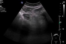
Instrumental diagnostics primarily involves ultrasound examination. Empyema of the gallbladder can manifest itself in different variations of the echographic picture. The most common ultrasound signs include intense and sometimes uneven structural disorders, altered echogenicity and thickness of the organ walls - both around the perimeter and locally. An enlarged gallbladder and pericystic fluid accumulation are detected. Bile is heterogeneous, may contain flakes, sediment and gaseous bubbles. [ 14 ]
When performing an ultrasound, it is necessary to take into account that the echo picture in case of gallbladder empyema can change quite quickly. A standard examination is performed using a convex sensor. After the procedure, the doctor fills out a diagnostic protocol, in which he describes all the parameters and changes in the gallbladder seen (position, shape, size, condition of the walls, inclusions, contents in the lumen, condition of the surrounding tissue).
As for endoscopic examination – in particular, retrograde cholangiopancreatography – if empyema is suspected, it is not performed in order not to waste time and to begin surgical treatment as soon as possible.
Additionally, an X-ray examination may be prescribed, which consists of a survey X-ray of the right hypochondrium, intravenous cholecystography. Less often, magnetic resonance imaging is used, which allows obtaining a direct picture of the biliary system and pancreatic ducts.
Differential diagnosis
Empyema of the gallbladder should first of all be distinguished from dropsy of the same organ. Dropsy develops as a result of complete or partial obstruction of the gallbladder duct, as a result of which mucus and exudate accumulate in the lumen of the gallbladder. Dropsy occurs after the cessation of bile outflow. The main characteristics of the pathology are occlusion of the gallbladder neck or duct by a calculus against the background of low virulence of the bacterial flora. The gallbladder absorbs the components of bile, microbes die, the contents of the gallbladder become discolored and mucous. During a physical examination of patients, it is possible to palpate an enlarged, stretched, painless gallbladder and its bottom. In a virulent infection, the gallbladder walls are thickened, and pus forms in the cavity.
The main method of differential diagnostics remains ultrasound examination. In the lumen of the organ, dense echo structures are examined that can move when changing the position of the body. Ultrasound transmits fairly reliable information - about 96-98%.
Auxiliary differential diagnostics are carried out with perforated ulcer, acute appendicitis, acute intestinal obstruction, right-sided pneumonia, urolithiasis, myocardial infarction (cholecystocardial syndrome), as well as with cholangitis, gangrenous or purulent cholecystitis.
To exclude diseases with similar clinical presentations, it is possible to use the following differential diagnostic methods:
- liver function tests;
- measurements of pancreatic enzyme levels;
- abdominal ultrasonography;
- tests with cholecystokinin, etc.
Who to contact?
Treatment gallbladder empyema
The main components of treatment for gallbladder empyema are urgent surgical decompression measures and cholecystectomy. Prescribing medications is an auxiliary method, including antibiotic therapy.
Basic treatment areas:
- prevention of complications in the form of perforation, etc.;
- unconditional removal of an organ.
The first stage of treatment is emergency decompression of the gallbladder, which is necessary to reduce the degree of compression of the surrounding tissues. If the patient has hemodynamic instability or there are contraindications to surgery (concomitant severe pathologies), then you can use the opportunity to perform hepatic drainage of the gallbladder under X-ray control, the essence of which is to remove exudate and pus from the organ. This procedure will allow decompression of the bile ducts, which will lead to a quick and significant improvement in the patient's well-being. However, such a measure cannot guarantee a complete victory over the pathology and the prevention of septic complications. Given this, if there are no contraindications to surgery, it is imperative to perform cholecystectomy - but only after stabilization of hemodynamic parameters.
After surgery and gallbladder removal, it is important to carry out supportive treatment, including antibiotic therapy. This stage should continue until the temperature indicators are normalized and the level of leukocytes in the blood is stabilized. Antibiotics are prescribed based on the results of the study of antibiotic resistance of the culture, inoculated from the bile secretion. [ 15 ]
Further management of patients includes adherence to a rational diet, physical activity, and sanitation of infection foci. Outpatient observation, subsequent sanatorium and resort therapy, and psychological rehabilitation measures play an important role.
Medicines
Drug therapy begins immediately after the surgical intervention, which involves the removal of the gallbladder. Such treatment may include the following measures:
- Infusion therapy to eliminate intoxication and restore water-electrolyte and energy deficit.
- Antibacterial therapy:
- Ciprofloxacin orally 500-750 mg twice a day for ten days.
- Doxycycline orally or intravenously by drip: 200 mg/day is used on the first day, then 100-200 mg/day, depending on the severity of the condition, for two weeks.
- Erythromycin orally, on the first day - 400-600 mg, then - 200-400 mg every six hours. The duration of administration can be from one to two weeks. Tablets are taken between meals.
To avoid adverse effects and side effects during antibiotic therapy (dysbacteriosis, mycosis), an oral solution of Intraconazole is prescribed in the amount of 400 mg/day for ten days.
- Oral cephalosporins – for example, Cefuroxime 250-500 mg twice daily after meals for two weeks.
- Symptomatic drugs are used according to indications:
- Cisapride (a gastroprokinetic that increases the motility of the upper gastrointestinal tract) is taken at 10 mg up to 4 times a day, or Debridate at 100-200 mg up to 4 times a day, or Meteospasmil at 1 capsule three times a day, for at least two weeks.
- Hofitol 2 tablets three times a day before meals, or Allochol 2 tablets up to 4 times a day after meals, for at least a month.
- Polyenzyme preparations, 1-2 doses before meals for three weeks, for several weeks.
- Antacids, one dose 1.5-2 hours after meals.
- Painkillers, antispasmodic drugs, depending on the required clinical effect.
Among the possible side effects of treatment, the most common are stool instability, abdominal pain, skin itching, and increased gas formation. Such symptoms require correction of both medications and diet.
Surgical treatment
Cholecystectomy is a surgical procedure that involves removing the gallbladder, an organ that stores bile that is produced in the liver and is involved in the digestive process.
Cholecystectomy is a mandatory treatment method for the development of gallbladder empyema, and the operation must be urgent to prevent the occurrence of life-threatening complications. In recent years, the intervention has been performed mainly laparoscopically, using a laparoscope (a special device with a video camera) and specific instruments. [ 16 ]
Laparoscopic cholecystectomy is rarely accompanied by complications, although in rare cases the likelihood of their development remains. Among the possible complications are the following:
- bleeding, blood clots;
- problems with the cardiovascular system;
- infection;
- damage to nearby organs (eg, small intestine, liver);
- pancreatitis;
- pneumonia.
The degree of risk of complications depends largely on the general health of the person and the initial causes of the development of acute cholecystitis.
Preparation for surgery includes the following points:
- assessment of hematological parameters and the state of vital organs;
- stabilization of hematological parameters.
All preparatory activities must be carried out within no more than two hours.
Cholecystectomy is performed using general anesthesia (intravenous). The operation itself is performed using a minimally invasive laparoscopic or traditional open method.
During laparoscopic surgery, the surgeon makes 2-4 punctures in the abdominal wall. A special tube equipped with a video camera is inserted into one of the punctures: the doctor has the opportunity to look at the monitor installed in the operating room and control the surgical instruments inserted through the remaining punctures into the abdominal cavity. Laparoscopic gallbladder removal lasts about 1.5-2 hours.
Sometimes, laparoscopy may not be possible, and the surgeon has to perform the operation using open access. The procedure is as follows. In the right segment of the abdominal cavity, closer to the costal arch, the doctor makes an incision measuring 3-10 cm, lifts the tissue to release the liver, and then removes the gallbladder. After control cholangiography, sutures are applied. The duration of open cholecystectomy is one and a half to two hours. [ 17 ]
The patient remains in the operating room or in the intensive care unit until the anesthesia wears off. Then he is transferred to a regular ward, where further recovery takes place.
After laparoscopic cholecystectomy, the patient may be released home on the third or fourth day, depending on his condition. Indications for discharge are as follows: the patient can eat and drink, move independently, with satisfactory general health and no complications.
After open cholecystectomy, the patient remains in hospital for a little longer until adequate recovery.
The postoperative period after cholecystectomy associated with gallbladder empyema is necessarily accompanied by antibiotic therapy. Antibiotics are prescribed until the leukocyte count in the blood stabilizes: at first, antibacterial agents are administered by intravenous infusions, then they switch to taking the drugs orally.
In the first few days, the patient is recommended to stay in bed, but the patient should periodically try to get up, which is necessary to prevent postoperative complications (such as pneumonia, adhesions, etc.). Until the gases pass, eating is prohibited: usually gases begin to pass 24-48 hours after the operation. Then you can eat little by little, starting with pureed soups, liquid mashed potatoes in water. After some time, liquid porridges, pureed vegetables and meat are introduced into the diet.
Prevention
Acute cholecystitis, which is complicated by empyema of the gallbladder, is one of the most common diseases of the gastrointestinal tract. Therefore, preventive measures, first of all, must be aimed at preventing the development of an inflammatory disease of the organ. Thus, the occurrence of acute cholecystitis is most often provoked by infection. Infectious agents enter the gallbladder in several ways:
- with blood;
- from the intestines;
- through the vessels of the lymphatic system.
With the lymph and blood flow, the infection penetrates the bladder if there are violations of the protective function of the liver. If there are failures of the motor function of the bile duct, then microbes can enter from the intestine. The inflammatory process develops against the background of a violation of the motor function of the bladder and bile retention.
Bile stagnation is caused by the presence of stones, elongation and tortuosity of the cystic duct, or its narrowing. In cholelithiasis, the incidence of acute inflammatory process is up to 90%. Due to the blockage of the duct by the stone, the entry of bile into the intestine becomes impossible, as a result, intravesical pressure increases, the walls stretch, blood circulation is disrupted, which leads to the onset of an inflammatory reaction.
What can be done to reduce the risk of developing acute cholecystitis and empyema of the gallbladder? Doctors give the following recommendations:
- eat fractionally, 5-6 times a day, without overeating or periods of fasting;
- avoid fatty, fried, salty, and overly spicy foods;
- get rid of bad habits such as smoking and drinking alcohol;
- lead an active lifestyle (a sedentary lifestyle contributes to the formation of stagnation);
- monitor your body weight and prevent the development of obesity.
Here are some foods that are recommended to be excluded from the diet, especially in cases where there are risk factors for the development of gallbladder empyema:
- fried, spicy, salty, too sour and fatty foods;
- hot sauces and seasonings (including mayonnaise, adjika, mustard, horseradish);
- heavy cream and sour cream, a large amount of butter;
- beans, peas;
- coffee, alcoholic drinks, cocoa, soda;
- chocolate, candy, baked goods;
- sour fruits, coarse-fibered vegetables.
It is important to promptly treat any pathologies of the digestive tract, infections of the genitourinary system, diseases of the ENT organs. If suspicious symptoms appear, it is necessary to contact a doctor as soon as possible.
Forecast
Empyema of the gallbladder can be fatal if the patient does not receive timely medical care and surgery. A good prognosis can only be spoken of if the pathology was detected in time and the patient did not have perforation, necrotic and septic complications. With the development of peritonitis and generalized sepsis, the prognosis worsens sharply.
In general, the outcome of the pathology often depends on the patient’s age and general health.
Timely therapy with its early start ensures a favorable prognosis: treatment ends with the patient’s complete recovery and return to his usual active activity. [ 18 ]
Patients belonging to the elderly and senile age category, as well as patients with immunodeficiency conditions and severe concomitant pathologies (for example, with decompensated diabetes mellitus) belong to a special risk group: progressive empyema in such patients can activate the development of septic complications, which are complex conditions that pose a threat to life. In addition, severe stretching and atrophic processes in the walls of the organ can cause their rupture (perforation), with the subsequent formation of bile peritonitis.
There is also some risk in the form of postoperative complications: operated empyema of the gallbladder can be complicated by wound infection, bleeding, and the development of a subhepatic abscess. However, timely medical care in the form of competent surgical and subsequent restorative treatment allows making the prognosis of the disease favorable.