Medical expert of the article

New publications
Dry eyes (dry eye syndrome)
Last reviewed: 04.07.2025

All iLive content is medically reviewed or fact checked to ensure as much factual accuracy as possible.
We have strict sourcing guidelines and only link to reputable media sites, academic research institutions and, whenever possible, medically peer reviewed studies. Note that the numbers in parentheses ([1], [2], etc.) are clickable links to these studies.
If you feel that any of our content is inaccurate, out-of-date, or otherwise questionable, please select it and press Ctrl + Enter.
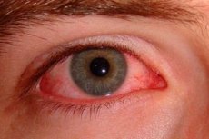
Dry eyes (Sjogren's syndrome) is a chronic disease with primary damage to the lacrimal and salivary glands. Dry eyes syndrome develops slowly and is chronic with periods of remission and exacerbation due to a lack of lacrimal fluid entering the conjunctival sac to moisten the anterior wall of the eyeball. As a result, periodic drying of the conjunctiva and cornea occurs, which leads to an unpleasant sensation of dryness, burning, itching and a sensation of a foreign body under the eyelids, photophobia, poor tolerance of wind and smoke. All these symptoms of dry eyes worsen in the evening.
Causes dry eyes
The causes of dry eyes are unknown. Some patients show signs of rheumatoid arthritis or other symptoms of connective tissue damage. Women over 40 years of age are more likely to get sick (90%), usually with the onset of menopause.
Symptoms dry eyes
Dry eyes have the following symptoms - irritation, foreign body sensation, burning, mucous thread-like discharge and periodic "fogging". Less common symptoms of dry eyes are itching, photophobia and tiredness or a feeling of heaviness in the eyes. Patients with filamentous keratin may complain of severe pain when blinking. Patients rarely complain of dry eyes, although some may note a lack of emotional tears or an inadequate reaction of tear secretion to an irritant (for example, onions). Dry eye symptoms are often aggravated by external factors associated with increased tear evaporation (for example, wind, air conditioning, central heating) or by very long reading, when the frequency of blinking movements is significantly reduced. Dry eye symptoms are also relieved by closing the eyes.
Disorders of the tear film
An early sign of dry eyes is mucin threads. Normally, when the tear film breaks, the mucin layer mixes with the lipid layer, but is quickly washed away. In a "dry" eye, mucin mixed with the lipid layer begins to accumulate in the tear film and shifts when blinking. A funny thing about mucin is that it dries out very quickly and rehydrates very slowly.
Marginal tear meniscus is a unit of measurement of the volume of the aqueous layer in the tear film. Normally, the volume of the meniscus fluctuates in height from 0.1 to 0.5 mm and forms a convex strip with a regular upper edge. In dry eyes, the meniscus may acquire a concave shape, become uneven, thin, or absent.
Foamy discharge in the tear film or along the edge of the eyelid is observed when the function of the meibomian glands is impaired.
Keratopathy
Punctate epitheliopathy affects the lower half of the cornea.
Corneal filaments consist of small, comma-shaped lumps of mucus at the level of the epithelium, attached at one end to the surface of the cornea; the free end moves with blinking.
Filamentous infiltrates are translucent, white-gray, slightly protruding formations of various sizes and shapes. They consist of mucus, epithelioid cells, and protein-lipid components. They are usually detected together with mucous threads when stained with rose bengal.
It is important to remember that dry eye contributes to the development of bacterial keratitis and frequent ulceration, which can lead to perforation.
Stages
There are 3 stages of eye damage: hyposecretion of lacrimal fluid, dry conjunctivitis, dry keratoconjunctivitis. Due to eye irritation at the first stages of the disease, lacrimation reflexively increases, which can be accompanied by a clinical picture of hypersecretion of tears - stagnation of tears and even lacrimation. Later, the secretion of tears with eye irritation sharply decreases, and there are no tears when crying. A viscous threadlike secretion consisting of tears and exfoliating epithelial cells is found in the conjunctival sac. The conjunctiva is moderately hyperemic, papillary hypertrophy is often observed along the upper edge of the cartilage. Superficial, small opacities of various sizes and shapes, stained with fluorscein, initially appear in the lower half of the cornea, and later - throughout the cornea. "Dry eyes" tend to progress, and other organs and systems of the body may be affected: dryness of the oral mucosa, nasopharynx, genitals, chronic polyarthritis, and later - disorders of the liver, intestines, cardiovascular system and genitourinary organs.
 [ 7 ]
[ 7 ]
Diagnostics dry eyes
When diagnosing dry eyes, it is necessary to take into account the patient's characteristic complaints, the results of a biomicroscopic examination of the edges of the eyelids, conjunctiva and cornea, as well as specific tests.
Special tests for dry eyes
- Norm's test - a test that evaluates the stability of the tear film. When looking down with the eyelid pulled back, a 0.1-0.2% fluorescein solution is instilled into the limbus area for 12 hours. After turning on the slit lamp, the patient should not blink. A tear film breakup time of less than 10 seconds is of diagnostic value.
- Schirmer's test with a standard strip of filter paper, one end of which is inserted behind the lower eyelid. After 5 minutes, the strip is removed and the length of the moistened part is measured: its value of less than 10 mm may indicate a slight decrease in the secretion of tear fluid, and less than 5 mm - a significant one.
- A test with a 1% solution of Rose Bengal is particularly informative, as it allows one to identify dead (stained) epithelial cells that cover the cornea and conjunctiva.
Diagnosis of dry eyes is associated with some difficulties and is based only on the results of a comprehensive assessment of the patient's complaints and symptoms, as well as the results of functional tests.
Tear film breakup time
The tear film breakup time is an indicator of its stability. It is measured as follows:
- fluorescein is instilled into the lower conjunctival fornix;
- the patient is asked to blink several times and then not blink;
- The tear film is examined in a wide section of a slit lamp with a cobalt blue filter. After some time, tears in the tear film can be seen, indicating the formation of dry areas.
The time between the last blink and the appearance of the first randomly located dry areas is taken into account. Their appearance always in one place should not be taken into account, since this is not caused by instability of the tear film, but is a local feature of the corneal relief. The time of appearance of dry areas in less than 10 seconds is a deviation from the norm.
Bengal pink
It is used to stain non-viable epithelial cells and mucin. Bengal rose stains the altered bulbar conjunctiva in the form of two triangles with their bases towards the limbus. Corneal filaments and infiltrates are also stained, but more intensely. The disadvantage of Bengal rose is that it can cause prolonged irritation of the eye, especially with pronounced "dry" eye. To reduce irritation, a small number of drops can be used, however, it is better not to use local anesthetics before instillation, as they can cause a false positive result.
[ 12 ], [ 13 ], [ 14 ], [ 15 ]
Schirmer test
It is used when a deficiency of tear fluid is suspected without biomicroscopic signs of dry eye. The test involves measuring the moistened portion of special paper filters 5 mm wide and 35 mm long (No. 41 Whatman). The test can be performed with or without local anesthesia. When performing the test without anesthesia (Schirmer 1), total, primary and reflex tear production are measured, and with the use of an anesthetic (Schirmer 2), only primary secretion is measured. In practice, local anesthesia reduces reflex secretion, but does not eliminate it completely. The test is performed as follows:
- carefully remove any existing tears;
- a paper filter, bent at a distance of 5 mm from one end, is placed in the conjunctival cavity between the middle third and the outer third of the lower eyelid, without touching the cornea;
- the patient is asked to keep their eyes open and blink as usual;
- After 5 minutes, the filters are removed and the amount of moisture is assessed.
The normal result is more than 15 mm without anesthesia and slightly less with anesthesia. The range between 6 and 10 mm is the normal range, and a result less than 6 mm indicates decreased secretion.
What do need to examine?
How to examine?
Who to contact?
Treatment dry eyes
Dry eye treatment is very difficult. Individual selection of medications is necessary.
Recommended by:
- constant instillation of artificial tears;
- at night, prescribe a disinfectant ointment or eye gel Solcoseryl or Actovegin;
- eliminate the cause that caused "dry eyes" (treatment of the underlying disease);
- avoid staying in dry and hot rooms for a long time;
If necessary, special obturators are introduced into the lacrimal canals or the lacrimal points are occluded using surgical methods.