Medical expert of the article

New publications
Diagnosis of the pancreas
Last reviewed: 05.07.2025

All iLive content is medically reviewed or fact checked to ensure as much factual accuracy as possible.
We have strict sourcing guidelines and only link to reputable media sites, academic research institutions and, whenever possible, medically peer reviewed studies. Note that the numbers in parentheses ([1], [2], etc.) are clickable links to these studies.
If you feel that any of our content is inaccurate, out-of-date, or otherwise questionable, please select it and press Ctrl + Enter.
Patients with pancreatic diseases may complain of abdominal pain, as well as dyspeptic symptoms and general weakness.
Complaints
Abdominal pain, quite varied in duration and character, is localized most often in the upper half of the abdomen, mainly in the epigastric region or left hypochondrium, radiating to the back. They can be sharp, intense, encircling in nature, with irradiation to the lumbar region, which in acute pancreatitis is associated with a violation of the outflow of secretion from the pancreas and the effect of its own proteolytic enzymes. Prolonged and intense pain is characteristic of tumors; they often intensify when the patient is lying on his back, which forces patients to take a semi-bent position.
Dyspeptic symptoms, nausea, and vomiting often occur in various diseases of the pancreas as a result of changes in its enzymatic activity or reflexively.
Mechanical jaundice with itching of the skin is typical for damage to the head of the pancreas with a violation of the outflow of bile.
 [ 1 ], [ 2 ], [ 3 ], [ 4 ], [ 5 ]
Physical methods of examination of the pancreas
Examination reveals exhaustion, jaundice with its characteristic consequences in the form of scratching, hemorrhage. Palpation of the pancreas remains an ineffective method. Only with a pronounced increase in the gland due to tumor damage, with deep sliding palpation is it possible to detect a neoplasm.
Additional methods of examination of the pancreas
Laboratory and instrumental methods allow more accurate detection of the active destructive process in the gland; assessment of the residual exocrine pancreatic function; assessment of the endocrine function of the pancreas and assessment of the morphological features of the gland.
X-ray examination of the pancreas. A general X-ray of the abdominal cavity allows detecting calcifications in the gland. When introducing barium contrast, it is possible to obtain indirect signs of inflammatory and neoplastic processes in the proximal part of the pancreas, taking into account changes in the mucous membrane of the duodenum.
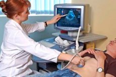
Ultrasound examination of the pancreas. This non-invasive and safe method allows detecting cysts and neoplasms in the pancreas, as well as determining its enlargement as a result of edema or compaction (fibrosis).
Computer tomography. In patients with obesity and intestinal obstruction, ultrasound examination is usually difficult, so it is advisable to conduct a computer tomography, which also allows you to evaluate morphological changes in the gland, identify a tumor, cyst, foci of inflammation, edema.
Angiography of pancreatic arteries. Selective angiography of the arteries supplying blood to the pancreas is useful in diagnosing tumors. It allows for detection of narrowing of the lumen of the vessels and their abnormal position. This examination is usually performed after ultrasound and computed tomography.
Endoscopic retrograde cholangiopancreatography. This study is considered one of the most valuable methods of visualizing the pancreatic and bile ducts. An iodinated contrast agent is introduced into the common bile duct through an endoscope, and then an X-ray is taken, which allows not only to establish the cause of mechanical jaundice, but also to identify changes in the pancreas characteristic of inflammatory and neoplastic processes. In chronic pancreatitis, the duct may be deformed, with areas of narrowing and widening visible. In the presence of a tumor, isolated stenosis of the duct or its complete obstruction is possible.
Radioisotope pancreas imaging. This is a pancreas imaging test using methionine labeled with a radioactive isotope of selenium and is generally much less accurate than the other imaging methods listed above.
Pancreatic enzymes in blood and urine. Pancreatic tissue necrosis due to pancreatic duct obstruction can be assessed by elevated pancreatic enzyme concentrations in the blood, urine, and other body fluids. The most common measurements are amylase and lipase activity. During acute pancreatitis, elevated serum amylase levels persist for up to 10 days and are usually accompanied by hyperamylasuria. Increased serum and urine amylase levels occur not only in pancreatitis, but also in biliary tract pathology, gastric ulcer perforation, intestinal obstruction, and some viral diseases, which is apparently associated with concomitant pancreatic damage.
Since amylase enters the blood not only from the pancreas but also from the salivary glands, attempts are currently being made to determine its isoenzymes. Using radioimmune research, the activity of other enzymes in the blood serum is assessed - trypsin, lipase, elastase.
Pancreatic function testing. The exocrine function of the pancreas is assessed using direct and indirect stimulation. Direct stimulation consists of parenteral administration of a number of hormones, in particular secretin and cholecystokinin, as well as their combination. Indirect stimulation consists of oral administration of nutrients. In both cases, pancreatic enzymes are measured - amylase, trypsin, lipase (the concentration of which, under the influence of secretin, initially decreases slightly and then increases) in the duodenal contents, which are obtained using a probe. An additional and important method for assessing the exocrine function of the pancreas is fecal testing to determine the content of fats and protein products.
Quantitative assessment of the fat content in feces, as well as chymotrypsin and trypsin, allows us to detect a progressive decline in gland function quite accurately.
The glucose tolerance test allows us to assess the endocrine function of the pancreas, which is impaired in 3/4 of patients with pancreatitis or pancreatic tumors.
The study of pancreatic function, especially the exocrine function, is important in patients with malabsorption to clarify the cause of this pathology and, in particular, to determine the role of decreased pancreatic function.
Who to contact?