Medical expert of the article

New publications
Dangerous moles: symptoms, how to recognize, treatment, prognosis
Last reviewed: 04.07.2025

All iLive content is medically reviewed or fact checked to ensure as much factual accuracy as possible.
We have strict sourcing guidelines and only link to reputable media sites, academic research institutions and, whenever possible, medically peer reviewed studies. Note that the numbers in parentheses ([1], [2], etc.) are clickable links to these studies.
If you feel that any of our content is inaccurate, out-of-date, or otherwise questionable, please select it and press Ctrl + Enter.
In medical terminology, a mole is called a “nevus” (from the Latin “naevus maternus”) – it is a formation on the human body that consists of cells that produce the pigment melanin.
The presence of moles should not cause concern, however, some dangerous moles can provoke serious oncological diseases. To avoid possible unpleasant problems, you need to carefully monitor them. The transformation of an ordinary mole into a dangerous malignant tumor is no longer a cosmetic problem, but a serious oncological disease. Thanks to the achievements of modern cosmetology and medicine, today there are many ways to avoid unpleasant consequences.
 [ 1 ]
[ 1 ]
Causes of a dangerous mole
Many superstitious people think that the number of moles determines a person's fate. The more moles, the happier the person. Doctors have a different opinion on this matter, since dangerous moles on the human body create big problems, sometimes degenerating into malignant melanoma or basal cell skin cancer. Let's list the main reasons why ordinary moles degenerate into dangerous ones. These are:
- Skin development defects. Such causes usually go unnoticed at birth, and are identified in the third or fourth year of a child's life, when dangerous birthmarks noticeably increase in size.
- Heredity. The fact that moles are inherited was noticed long before the advent of DNA tests. Some neoplasms are encoded in the DNA molecule by a chain of genes that is passed from parents to children. However, acquired moles are not inherited.
- Ultraviolet in large quantities. During tanning, melanin is produced in the basal layer of the skin by special cells called melanocytes.
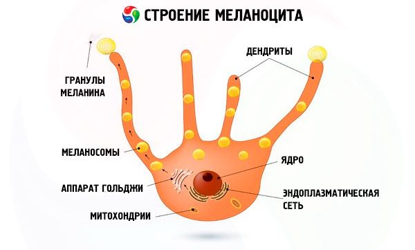
- With strong exposure to ultraviolet rays, melanotropic hormone is produced, which, in turn, increases the number of melanocytes. In other words, instead of tanning, an intensive increase in melanocytes begins. Such moles (nevi) are called acquired. Therefore, exposure to active sun should be limited. Also, passion for solarium is not recommended. Statistics show that people with sensitive and naturally light skin, with a large number of pigmented, birthmarks and freckles, as well as women over 30, are especially susceptible to ultraviolet radiation.
- Trauma. Any scratches, small wounds, insect bites, as well as self-removal of a mole, hair pulling, etc. can provoke the development of dangerous moles. The fact is that mechanical damage affects different layers of the skin, therefore, tissue inflammation occurs, and biologically active substances are produced that stimulate cell growth.
- Hormonal risk factors. Most often, the hormonal trigger for the development of moles is the melanotropic hormone of the pituitary gland. There are pathological and physiological hormonal changes in the body, which can serve as a background for the development of dangerous moles: adolescence during puberty, during pregnancy and in people prone to the development of endocrinological diseases. In this case, we are talking about.
- Bacterial and viral infections. Recently, this version has been considered in medicine. The mechanism of nevi appearance is similar to injuries, as a result of which, against the background of the inflammatory process, a neoplasm develops.
Based on the above, we can conclude that the risk group includes patients with acquired moles (nevi), moreover, acquired neoplasms have a greater tendency to degenerate into dangerous moles.
Pathogenesis
Symptoms of a dangerous mole
Benign moles do not pose a danger to human health. However, a common mole can become dangerous and turn into a malignant tumor.
First signs
Dangerous moles (nevi) on the body can turn into malignant formations. To prevent this, you should inspect your body once a month, assessing the condition of your moles. Particular attention should be paid to moles located in hard-to-reach places, such as the back, head, and there are cases of moles localized on the mucous membrane of the body. In such situations, you should seek help from a loved one. To identify dangerous moles, you need to know the first signs by which you can determine them. Let's list some of them:
- Pay attention to the color of the mole. Its color should be uniform. The color may be similar to the skin, or vice versa, the color of the mole changes radically to the opposite, black color. There are cases when the edges are unevenly colored, that is, one part of the edge is light, gradually turning into a darker tone. If the mole has a change in color, or it can have a mixed color - this is the first sign of a malignant tumor.
- Active growth is observed, the mole increases greatly in size. At the same time, it can become very dense. Normal mole sizes are from 0.6 to 1 cm. Sometimes the mole decreases. If the parameters exceed the permissible limits, you should immediately consult a specialist.
- Hyperemia (swelling) may form around the mole, which spreads into the epidermal tissue structures.
- The marked edges of the mole become pale and more blurred.
- Hair loss from the surface of a mole.
- Feeling of pain, itching. The mole may itch, prick, bleed, forming a dense crust on the surface.
- The mole changes its configuration, the edges become blurred, and asymmetry is observed.
- Sometimes the lymph nodes become enlarged.
Dangerous moles (including melanoma) can spread not only throughout the body, but also deep into the tissues, with metastases affecting literally all organs, including the liver, kidneys, lungs, etc.
Dangerous birthmarks in children
Immediately after birth, mothers examine their baby's body, and many of them are concerned about the problem of moles. The process of appearance and formation of nevi occurs up to 25 years, however, there are congenital moles. Real moles on the child's body appear after six years. With age, their number can increase or decrease. Children's moles are no different from moles of an adult. They can be convex, flat, light brown in color, up to 1 cm in diameter, but mostly these are small pigment spots that are difficult to determine by touch. According to statistics, in 80% of cases, moles in a child do not cause problems, but this does not mean that parents should not monitor their condition. Dangerous moles on a child's body detected in a timely manner can reveal a serious oncological disease in its early stages.
Small moles in children are absolutely safe. Large moles and pigment spots are at risk. According to medical statistics, about 40% of them develop into a dangerous malignant tumor. Dangerous moles on a child's body are:
- large moles;
- the appearance of a large number of moles;
- moles that are located in an easily accessible place and can be easily injured.
Therefore, such formations on the child's body should be kept under close attention. Also, if a mole on the child's body itches, peels, changes its structure, shape or color, it is necessary to immediately contact specialists.
Dangerous birthmarks in children can be divided into several types. These are:
- Red spots. The most common formations on the baby's body. They are formed as a result of direct contact of the fetus with the pelvic bones of the expectant mother. Such spots disappear on their own within a year.
- Brown pigment spots. These are common moles that may disappear and reappear over time. Such nevi often do not pose a particular danger, but their condition should be monitored.
- Port-wine stains. These are formations that are dilated capillaries located on the child's face or head. They tend to increase in size as the child grows older, but their color does not change. Laser treatment is recommended, starting from an early age.
- Strawberry hemangioma. A bright red, soft to the touch, convex birthmark that can be congenital or appear immediately after birth during the first weeks of life. The birthmark can grow, change shape, and color. Such a formation cannot be treated, moreover, if you start the removal procedure, there may be the most irreversible consequences.
- Cavernous hemangioma. A large cluster of deep blood vessels. It has a bluish-gray color. It disappears on its own by the age of 12.
- Pigmented nevus. A pigmented spot of relatively small size, can be congenital or form independently in early childhood. Popularly known as a "birthmark". It does not pose a particular danger, however, there are some forms, such as dysplastic nevi, which can degenerate into malignant skin tumors.
- Red birthmark (angioma). A reddish pigment spot located on any part of the child's skin. A benign formation of vascular origin. Angioma in children can be an emotional problem or a cosmetic defect. In rare cases, a malignant nature of the formation can be observed.
As you can see, not all types of the above nevi and birthmarks are dangerous, but if certain factors are present, they may be at risk. According to medical statistics, the probability of oncological formations in children increases 10 times.
[ 6 ]
Forms
Moles (nevi) are benign formations that by their nature do not cause any particular unpleasant sensations. However, there are cases when these completely unobtrusive black spots change, turning into dangerous moles. Therefore, it is necessary to periodically examine your body, carefully look at moles in order to prevent pathological changes.
Which moles are dangerous?
What causes a normal pigment spot to degenerate into malignant melanoma? The reasons may be as follows:
- the mole is in an easily accessible place, constantly "rubs" with clothes, and is easily injured when touched. Experts advise removing such nevi. Before starting the removal process, make sure that it is a benign formation;
- excessive use of solarium;
- active sun rays. Experts advise using sunscreens and covering moles with cotton clothing.
Only small congenital pigment spots can be absolutely safe. All other pigmented formations on the body can be safely questioned, so carefully monitor the changes that occur so as not to miss dangerous moles. The main thing is not to delay a visit to the doctor.
Red moles on the body are a signal of dangerous diseases
Red moles are called angiomas in medical terminology. This is a collection of small vessels and capillaries that are localized under the human skin, causing a change in color, in this case red predominates. Angiomas are a completely normal physiological phenomenon, but many believe that these are dangerous moles. Is this true?
Red moles can appear in large numbers. How can they be dangerous? Among the huge number of versions on this account, we can highlight some of them:
- liver function problem;
- insufficient amount of vitamin K (menadione) in the body;
- autoimmune diseases;
- oncological diseases;
- diseases of the gastrointestinal tract, especially pathology of the pancreas;
- hormonal changes (pregnancy, menopause, puberty).
- serious disruptions in the cardiovascular system of the body;
- poor nutrition, as a result of which a large amount of toxins accumulate in the intestines.
So, if six or more red moles appear on a small area of the body, this is already a signal of the development of a serious disease in the body. It would be a mistake to think that small red spots on the skin can be removed independently. This should not be done, as this can lead to bleeding, and subsequently cause cancer.
As a rule, red moles on the human body in limited quantities do not pose a particular danger, but if they quickly begin to spread, bother, or you notice other symptoms, this is already a serious signal that changes are occurring in the body. To date, there is no clear answer to the question of the appearance of red moles. The factors listed above can provoke their appearance.
In any case, if the above symptoms appear, you should immediately consult a doctor, because only a doctor can identify dangerous moles and determine which of them should be removed immediately.
Dangerous black moles
Black moles differ from others only in color. They, like all other moles, have a round shape, the correct size (there are small deviations), a smooth surface, etc. Basically, black moles appear equally on the body of men and women, and their color range is a feature of human skin. The color of a mole depends on the number of melanocytes, and not only. An important role is played by the melanotropic hormone, which is produced by the pituitary gland (the gland responsible for growth, development and metabolism in the human body), therefore, several body systems participate in the process of coloring moles.
The risk of a black mole transforming into melanoma is quite high. For example, dangerous moles can change their structure, and shades of gray or red can be added to the monotonous black color. This is a kind of signal that unfavorable processes are beginning to develop in the black mole. Therefore, it is necessary to monitor the dynamics of changes, which are expressed in the following:
- the surface must be smooth;
- clear symmetrical shape;
- no roughness or peeling;
- The mole should not bleed.
Doctors recommend paying special attention to large black moles with a diameter exceeding 6 mm.
Dangerous raised moles
Doctors consider convex moles to be the most vulnerable formation on the human body. They are dangerous because due to their large size and convexity they are constantly in the risk zone. At any moment you can feel contact with clothing or underwear, moreover, it is easy to catch and injure. The slightest injury to a mole is very dangerous, moreover, it can result in the development of oncological skin disease.
From a medical point of view, convex moles are less likely to transform into melanoma, as they are more visible and are always under control. However, it is advisable to remove convex moles.
[ 7 ]
Dangerous Large Moles
Large moles, especially those located on the face, always attract the attention of others.
Large moles are not always dangerous. If the nevus does not change its condition, there is no point in worrying. You just need to periodically monitor your appearance, undergo regular examinations. A large mole can become denser, hurt, itch, etc., in which case there is every reason to believe that it can transform into a malignant tumor.
Dangerous Flat Moles
Flat moles are called lentigo in medical terminology. This is the most common, harmless type of mole, the presence of which can go unnoticed. Flat moles are:
- solar;
- youthful;
- senile.
Solar moles are a consequence of the influence of ultraviolet rays on the skin. Their diameter is about 0.5 cm. The color can vary from light brown to black. The older the mole, the darker it is. Light-haired and fair-skinned people, as well as young people who are overly fond of solariums, are most susceptible to the appearance of lentigo. Solar moles are not dangerous to health and are not associated with chronic diseases.
Juvenile flat moles or juvenile lentigines appear on the skin of young people. These are dark spots in the form of a circle or oval with a diameter of 3 to 15 mm, which can also appear as a rash. The edges of juvenile lentigines can be jagged or simply smoothed. They can be localized not only on the skin, but also on the mucous membranes. The reason for the appearance of this form of moles is still unknown, but it can be definitely stated that this type of lentigo is formed by exposure to ultraviolet rays. Such moles can appear in childhood.
Senile flat moles appear in old age.
Flat moles are not dangerous, most often they are harmless to the body. You can get rid of flat moles with the help of bleaching creams and/or cosmetic lightening procedures. To reduce the risk of flat moles, you need to avoid ultraviolet rays, eat right and lead a healthy lifestyle.
Complications and consequences
There are no consequences from moles! There are consequences after the procedure of their removal, which depend on many reasons. Let's list some of them:
- characteristics of a mole, its parameters;
- qualifications, professional skills of a specialist;
- equipment, quality of medical products;
- the patient's body's reaction to the course of the operation.
After the operation, during the healing process, the wound is covered with a black crust, which should peel off on its own. Moreover, if the crust comes off prematurely, the wound will open again, into which infections and bacteria can penetrate, which can end in serious inflammation. The wound must be treated with a weak solution of manganese. During the healing period, the mole site must be protected from moisture, which means that you should avoid visiting the pool, sauna, or taking a bath.
In 2-3 weeks, a patch of fresh pink skin will appear instead of the black crust. The renewed patch of skin is particularly sensitive. It should be protected from ultraviolet rays, as pigment spots may appear in this area. Use sunscreen, cover the areas of the body with cotton clothing.
The remaining scars and marks gradually disappear, however, for a better effect they can be lubricated with cocoa butter, which is sold in pharmacies.
Complications
Problematic dangerous moles can provoke serious consequences and oncological diseases that really threaten a person's life. Experts recommend removing them if necessary. The following complications are possible after removing a mole:
- dull pain, burning, or itching;
- allergic reaction to anesthesia. To ensure that the mole removal does not go without complications, it is necessary to conduct a test for drug sensitivity before the procedure;
- the presence of scars and marks. For rapid healing, it is necessary to follow all the doctor's recommendations, use special ointments for healing and smoothing scars. Over time, these consequences disappear.
It is recommended to remove dangerous moles in a medical institution, entrusting this responsible process to a professional who has experience and knowledge. Visiting beauty salons is not advisable, as this can lead to serious negative consequences.
[ 10 ]
Is it dangerous to pick off a mole?
There is no point in removing moles without serious reasons. However, there are situations in which a mole is accidentally injured. This happens especially often if dangerous moles are on the wrist, neck, head, or come into contact with clothing. In any case, damage to moles can cause transformation into a malignant tumor.
If a mole is accidentally torn off, you need to stop the bleeding. Moisten a sterile swab with hydrogen peroxide and apply it to the damaged mole. Then, take a dry sterile swab and hold it for 15 minutes. After this, you must definitely consult an oncologist.
The most common cause of melanoma is trauma to a mole. Medical statistics show that about 40% of melanomas occur due to accidental trauma.
Is it dangerous to injure a mole when shaving?
Often dangerous moles are located in an easily accessible place on the face, and are injured during shaving. If the razor touches a mole, you need to stop the bleeding by treating the area with hydrogen peroxide. The materials used for treatment must be sterile.
If the razor has completely cut off the mole, it should be wrapped in a bandage or gauze soaked in saline solution and a histological analysis should be done. A partially injured mole should be shown to a dermatologist, who will then remove it and send it for analysis.
If a mole has been repeatedly slightly injured by a razor, it must be removed immediately. In addition, it is mandatory to undergo a histological examination of the excised tissue.
Diagnostics of a dangerous mole
To establish a correct diagnosis, the doctor must carefully examine dangerous moles and conduct the following types of examination:
- Collection of anamnesis. This type of diagnostics consists of asking the doctor the right questions to determine the family anamnesis. Detailed information is collected about which of the close relatives had dangerous moles or birthmarks, whether there was a diagnosed melanoma in the family. Next, the doctor should find out about the risk factors and causes that could cause changes in the nevus, as well as the presence of chronic pathology.
- Visual examination of moles - dermatoscopy. The presence of a malignant nature of a nevus can only be confirmed by a biopsy of a suspicious area of skin.
How to recognize a dangerous mole?
Any normal mole on the human body can change pathologically and become dangerous under the influence of certain reasons. In this case, it is important to be able to distinguish between its two states.
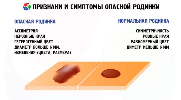
First of all, it is necessary to regularly pay attention to the appearance of the mole. Doctors have developed a special method that will help any interested person to recognize dangerous moles. The abbreviation of English letters ABCDE will indicate the key points to which special attention should be paid. By the way, this method is called ABCDE.
- A – asymmetry. The normal condition of a mole can be determined as follows. Visually divide the mole into two equal halves. If both halves are symmetrical to each other, there is no need to worry. If one half begins to grow to the side, you should sound the alarm.
- B – change in the edge of the mole. This is one of the signs of a malignant tumor – melanoma, when the edge of the mole becomes paler or blurry. In a normal state, a mole should have perfectly smooth edges.
- C – mixed color. By its nature, a mole always has one color. Heterogeneous color with non-uniform inclusions of other shades causes caution.
- D – diameter size. The mole should not exceed 6 mm in diameter. If this happens, consult a doctor immediately.
- E – change. In this case, any distortion of shape, size, color, etc. is implied.
If at least one symptom appears, immediately consult a specialist. The mole remains unchanged throughout life.
In order to make it easier to remember the above-mentioned signs, there is a Russian-language reminder called AKORD, each capital letter of this word corresponds to the first letter of dangerous symptoms:
- A - asymmetry;
- K - edge;
- O - color;
- P - size;
- D - dynamics.
Those at risk include those who have already had dangerous moles and had them removed, as well as those with hereditary factors for the development of neoplasms.
Tests
When examining and diagnosing nevi, no tests are required.
If dangerous moles are removed surgically, a general blood test, biochemical blood test, and urine test may be required. Such a list of tests is necessary for a general assessment of the body and internal organs. In this case, the tests reveal chronic diseases of the patient, which may subsequently affect the outcome of the disease. Sometimes the test results reveal contraindications to the removal of dangerous moles.
Instrumental diagnostics
Dangerous moles are diagnosed using digital dermatoscopy.
Using a dermatoscope, a clear digital image of a mole is obtained, which is displayed on a computer monitor. Thanks to the possibility of multiple optical magnification, the dermatologist analyzes in detail the shape, color, contours and other external signs of the mole. In addition, the procedure creates a "map" of moles located throughout the human body, which makes it possible to continue monitoring nevi for their further prevention.
Since nevi can be large and asymmetrical, we need data on when the pigmented lesion began to change. It is safe to say that any dangerous moles require a biopsy.
Biopsy is a reliable and accurate diagnostic method for determining malignant neoplasms, melanoma metastasis nevus. The accuracy of the biopsy method is equal to 100%, provided that the procedure itself is performed correctly.
There are two types of biopsy:
- puncture;
- total excisional;
During a puncture biopsy, a fragment of nevus tissue is taken using a special needle, the number of cells for analysis is limited. The procedure is performed under local anesthesia.
Total excisional biopsy is a diagnostic and therapeutic measure at the same time. This method removes the neoplasm and uses it for histological examination.
Histological examination is considered the most important and reliable. As a rule, this is the final type of diagnosis, in which a tissue fragment taken after a biopsy is assessed under a microscope.
If there is a suspicion of melanoma, in order to avoid the spread of metastases, ultrasound, X-ray, and MRI are additionally prescribed.
Differential diagnosis
All types of moles are differentiated from melanoma and basal cell skin cancer.
Who to contact?
Treatment of a dangerous mole
When starting treatment of moles, it is necessary to undergo diagnostics, preferably to have the results of tissue biopsy. Treatment of moles involves only their removal.
You can remove dangerous moles using the following methods:
- surgical removal of a mole;
- laser removal;
- cryotherapy;
- electrocoagulation;
- radio wave surgery.
Let's take a closer look at the action of these methods.
Surgical removal of moles. In some cases, dangerous moles are offered to be removed by surgical operation. In the event that the examination results indicate malignancy of the mole, the operation is performed only by an oncologist, after which additional radiation to the removal site and/or chemotherapy may be prescribed. A mole that does not have signs of malignancy can be removed by a dermatologist or cosmetologist.
The surgical method of removing a mole involves excising tissue with a scalpel. The pigment cells and the area of skin around the nevus are removed. The operation is performed under local anesthesia. A scar may remain after the operation. Recently, the surgical method for benign moles has not been used.
Laser removal of moles. The most popular, effective and sought-after method of removing moles in our time. During the removal procedure, the patient does not experience pain or discomfort. A slight tingling and warmth are felt. With the help of a laser, liquid is evaporated from the tissue, after which the cells die. The positive aspect of this method is the treatment of a large number of moles. The laser removal method does not leave scars. Large moles, the diameter of which reaches up to 2 cm, can be problematic. Sometimes there are cases when the procedure cannot be completed to the end and part of the mole remains untreated, subsequently it can grow again. In this case, the surgical method is more suitable.
Cryotherapy. A method of influencing a mole with a cryoapplicator treated with liquid nitrogen. Low temperature (-1960) destroys pathological tissue. The procedure is painless, allows the treated area to heal with minimal impact on the skin, leaving no traces at all.
Electrocoagulation. A fairly common method of removing moles. An electric current is used to affect the tissue. After the procedure, a tissue analysis can be done. After healing, there are practically no traces left.
Radio wave removal. The most effective method of removing nevi. Thermal energy generated from high-frequency waves cuts the tissue without affecting the mole. In this case, pigment cells evaporate, leaving a barely noticeable mark. The operation takes about 20 minutes. Side effects such as redness, swelling, inflammation are not observed.
Any of these methods of mole removal requires a highly qualified doctor who can accurately calculate the force of the procedure on the tissue of the mole. If the mole is not completely removed, it may appear again.
Medicines
A group of Russian scientists from the N. N. Petrov Oncology Research Institute (St. Petersburg) developed the drug "Refnot", which is successful in the treatment of disseminated forms of melanoma. The drug combines two biologically active components - tumor necrosis factor cytokinin and the hormone thymosin. In medical practice, Refont is used as an antitumor agent. The drug has been registered and approved for use.
The new generation of drugs "Ipilimumab" and "Nivolumab" stop the growth of cancer cells in the body for about 1 year. The combined use of drugs allows to reduce the size of the tumor at the last stage of the disease.
The drugs "Ipilimumab" and "Nivolumab" stimulate the immune system. Side effects of the drugs include chronic fatigue, diarrhea. In some cases, patients develop a rash.
Folk remedies
You can get rid of unwanted moles using folk medicine. Of course, compared to the surgical method, the treatment process with folk remedies is longer, but there are pros and cons. Folk treatment does not entail big financial problems, and primitive, mostly environmentally friendly food products or herbs are used for treatment.
In any case, if you decide to get rid of a mole using folk remedies, you must consult a dermatologist or oncologist to make sure the procedure is safe. The process of removing a mole in folk medicine is divided into two treatment methods.
The first method is to block the blood supply to the body of the mole by tying it with a thread at its base. This method of treatment has negative consequences, and the most harmless procedure can turn a mole into a dangerous melanoma. In the best case, the mole will disappear, and after some time it will appear again.
The second method of folk treatment is safer and involves using several recipes. Here are some of them.
- Vinegar essence, with the help of which mole cauterization is done. The procedure is performed once a day, after which the treated area is covered with a bandage to prevent infection. This is a rather painful procedure. The mole disappears within two weeks. If this does not happen, the procedure can be repeated in two or three weeks. According to folk healers, this is a very effective method of removing moles.
- Moles can be lubricated with linseed, castor oil, onion juice, lemon and garlic. Wiping with hydrogen peroxide, iodine, baking soda, natural honey is also popular. After such procedures, they significantly decrease in volume and then completely disappear.
- Removing moles and warts with an old grandmother's recipe. To do this, hard-boil 7 eggs, remove the yolks. Next, grind the dried pumpkin seeds into flour, after frying them, to get 5 tablespoons. Mix the yolks thoroughly with the pumpkin flour, and add 0.5 liters of vegetable oil. Stir in a glass container with a wooden spoon for thirty minutes. This completes the cooking process. The resulting medicine should be stored in the refrigerator. Take one tablespoon on an empty stomach in the morning for 1 week. After that, take a 5-day break and start taking it again until the medicine runs out.
- Cut a dry ear of grain and lightly prick a mole or wart with its sharp part. Then bury the straw in damp earth, and leave the ear itself on the surface. People say that when the straw rots, the mole or birthmark will disappear.
Removing moles using folk methods is characterized by the fact that as a result of treatment, no marks or scars remain on the body. The mole decreases in size and disappears without a trace.
Traditional methods of treatment will help to get rid of unwanted moles, but in any case, you need to remember that you need to diagnose and remove a mole only under the supervision of a doctor, even if it is an old, proven grandfather's method.
[ 13 ]
Herbal treatment
Nevi can be removed at home using medicinal herbs, many of which grow in the country or in the garden. For this, traditional healers use cauliflower juice, garlic, but the most common medicinal plant is celandine. To remove moles, use:
- The juice of the plant is applied directly to the mole 2-3 times a day, squeezing it directly from the stem and leaves of the plant.
- Celandine ointment, for the preparation of which it is necessary to mix young celandine leaves with baby cream or pork fat in a ratio of 1:2. Instead of leaves, you can use celandine juice, then you should take one part of juice and 4 parts of cream.
- Oil for lubricating moles, based on celandine. Grind dry leaves and pour them with vegetable oil. Put the container in a dark place for a week. After that, lubricate the nevus for one month 2-3 times a day.
- Celandine tincture. 100 grams of celandine leaves should be poured with 0.5 liters of alcohol and left for two weeks in a dark place. Then strain the tincture. Moles will disappear if you take the tincture 10-12 drops 3 times a day.
Good results in the treatment of dangerous moles are provided by external preparations. The most effective means used to treat moles and melanomas is the ointment "Stefalin", which contains only medicinal herbs. Stefalin painlessly removes moles and warts, leaving no scars. The procedure can be performed at home. The ointment has no side effects.
There is an opinion that the success of removing moles with medicinal herbs at home is a myth. Many people in their practice used the many years of experience of healers, as a result of which a positive effect was observed - the complete disappearance of moles
Prevention
Dangerous moles have a tendency to degenerate into melanoma. To prevent this from happening, doctors recommend following simple rules and recommendations for prevention, which boil down to the following:
- Check dangerous moles regularly according to the “AKORD melanoma” scheme. The recommended time for examination is early May and late September.
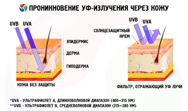
- Avoid sun exposure between 11 a.m. and 3 p.m. Use sunscreens to protect your skin from severe sunburn and reduce the risk of dangerous moles.
- Clothing should be loose, preferably made of cotton fabric.
- Never cover moles on your body with a plaster, as this creates a thermal effect that has a negative effect on the mole.
- Sunbathe only in the morning and evening.
- Dangerous moles cannot tolerate direct sunlight.
- Don't get carried away with visiting the solarium. This especially applies to women over 30.
- Use moisturizing creams.
- Monitor the condition of your skin. If you experience a rash, itching, or redness, consult a doctor. Such manifestations contribute to malignant formations of moles.
- Protect moles from mechanical damage. If a mole is in an inconvenient place and can be easily torn off, it is better to remove it. Regular injuries lead to inflammation and the development of melanoma.
- Chemicals used in everyday life can provoke dangerous moles to develop oncology. Some chemical compounds have a mutagenic effect on dangerous moles. Smoking can also be considered a risk factor.
- If you have dangerous moles on your body, you need to undergo preventive examination and visit a doctor approximately once a year.
People with fair and red hair and light skin are at risk, as they have a lower level of melanin in their bodies and are therefore more susceptible to the effects of sunlight.
[ 14 ]
Forecast
The prognosis of dangerous moles depends on the presence of signs of malignancy. An important criterion is the nature of the disease and the stage of its development.
The prognosis indicator for the development of melanoma depends on the number of skin layers involved in the oncological process. Thin melanomas, less than 1 cm in size, have good treatment efficiency indicators.
Moles without any signs of change have a favorable prognosis.